Copyright
©2012 Baishideng Publishing Group Co.
World J Gastroenterol. Apr 7, 2012; 18(13): 1459-1469
Published online Apr 7, 2012. doi: 10.3748/wjg.v18.i13.1459
Published online Apr 7, 2012. doi: 10.3748/wjg.v18.i13.1459
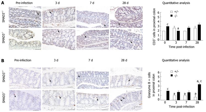
Figure 5 Immunohistochemical staining for (A) CD3 lymphocytes and (B) granzyme B in proximal colon tissue of drosophila mothers against decapentaplegic 3+/- and drosophila mothers against decapentaplegic-/- mice following infection with Helicobacter hepaticus.
aP < 0.05 vs baseline values; cP < 0.01 denotes significant interaction between genotypes [drosophila mothers against decapentaplegic (SMAD)3-/-vs SMAD3+/-] (n = 4-5 animals per stain). Arrows denote areas of positive staining. Scale bars represent 100 μm.
-
Citation: McCaskey SJ, Rondini EA, Clinthorne JF, Langohr IM, Gardner EM, Fenton JI. Increased presence of effector lymphocytes during
Helicobacter hepaticus -induced colitis. World J Gastroenterol 2012; 18(13): 1459-1469 - URL: https://www.wjgnet.com/1007-9327/full/v18/i13/1459.htm
- DOI: https://dx.doi.org/10.3748/wjg.v18.i13.1459


