Copyright
©2012 Baishideng Publishing Group Co.
World J Gastroenterol. Apr 7, 2012; 18(13): 1438-1447
Published online Apr 7, 2012. doi: 10.3748/wjg.v18.i13.1438
Published online Apr 7, 2012. doi: 10.3748/wjg.v18.i13.1438
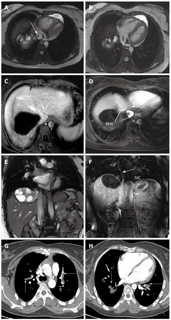
Figure 17 Hepatic hydatidosis in a 30-year-old woman who presented with short of breath, fatigue and edema to the lower limbs.
Magnetic resonance (MR) steady-state-free-precession sequences (A, B, E) and MR angiography (C, D, F) images showed the hydatid cyst invading the right hepatic vein (RHV), protruding in the inferior vein cava (IVC) (the bold white arrow) and in the right atrium (the white arrowhead). The mid hepatic vein (MHV) and the left hepatic vein (LHV) were normally patent (C). The multidetector computed tomography-angiography revealed diffuse pulmonary parasitic embolism (the thin white arrows) (G, H).
- Citation: Marrone G, Crino' F, Caruso S, Mamone G, Carollo V, Milazzo M, Gruttadauria S, Luca A, Gridelli B. Multidisciplinary imaging of liver hydatidosis. World J Gastroenterol 2012; 18(13): 1438-1447
- URL: https://www.wjgnet.com/1007-9327/full/v18/i13/1438.htm
- DOI: https://dx.doi.org/10.3748/wjg.v18.i13.1438


