Copyright
©2012 Baishideng Publishing Group Co.
World J Gastroenterol. Mar 28, 2012; 18(12): 1373-1378
Published online Mar 28, 2012. doi: 10.3748/wjg.v18.i12.1373
Published online Mar 28, 2012. doi: 10.3748/wjg.v18.i12.1373
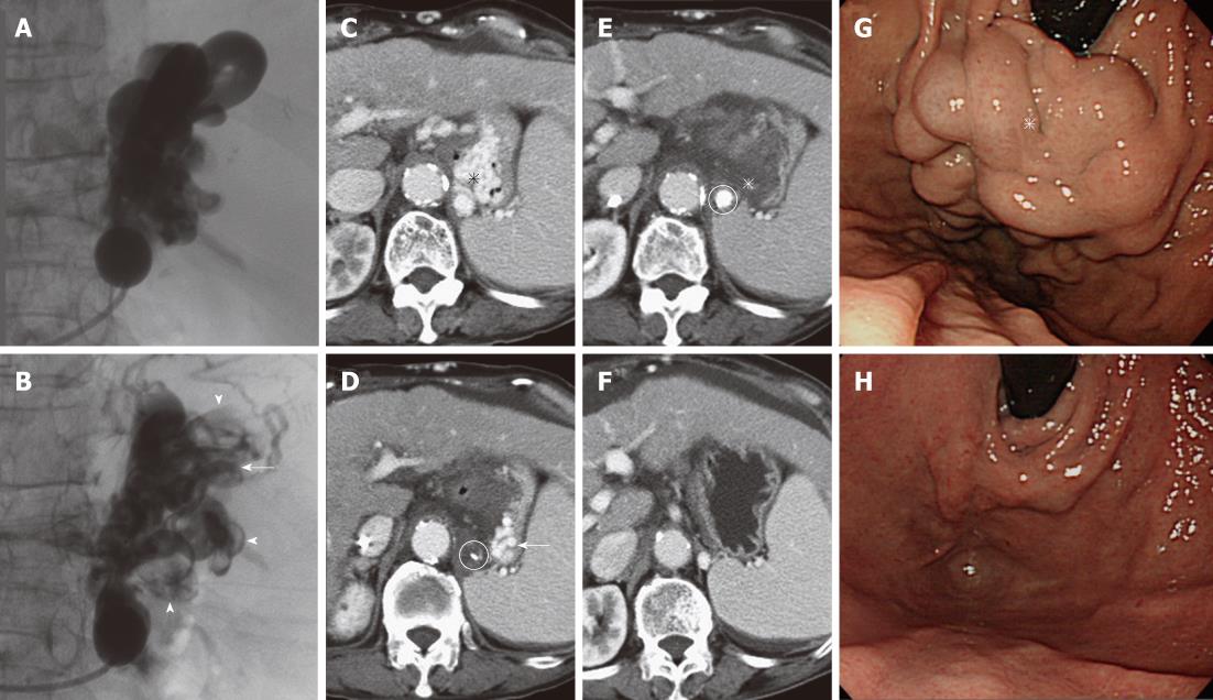
Figure 4 Additional injection of the sclerosant.
A: Fluoroscopic image obtained during the first balloon-occluded retrograde transvenous obliteration (BRTO) shows full opacification of the gastric varices and gastrorenal shunt; B: Fluoroscopic image obtained during the second BRTO (next day) shows partial opacification of the varices and shunt, suggesting residual varices (arrow) and thrombosis of the varices and shunt (arrowheads); C: Contrast-enhanced computed tomography (CE-CT) before BRTO shows large varices (asterisk); D: CE-CT after the first BRTO shows residual varices (arrow) in the lateral portion of the stomach. The microcatheter tip (circle) is in the gastrorenal shunt close to the varices; E: CE-CT after the second BRTO shows complete thrombosis of the varices (asterisk). The sclerosant with contrast medium (circle) is detected in the gastrorenal shunt; F: CE-CT 3 mo after BRTO shows complete disappearance of the varices; G: Endoscopy before BRTO shows bulky varices (asterisk); H: Endoscopy 3 mo after BRTO shows complete disappearance of the varices.
- Citation: Sonomura T, Ono W, Sato M, Sahara S, Nakata K, Sanda H, Kawai N, Minamiguchi H, Nakai M, Kishi K. Three benefits of microcatheters for retrograde transvenous obliteration of gastric varices. World J Gastroenterol 2012; 18(12): 1373-1378
- URL: https://www.wjgnet.com/1007-9327/full/v18/i12/1373.htm
- DOI: https://dx.doi.org/10.3748/wjg.v18.i12.1373


