Copyright
©2012 Baishideng Publishing Group Co.
World J Gastroenterol. Mar 28, 2012; 18(12): 1319-1327
Published online Mar 28, 2012. doi: 10.3748/wjg.v18.i12.1319
Published online Mar 28, 2012. doi: 10.3748/wjg.v18.i12.1319
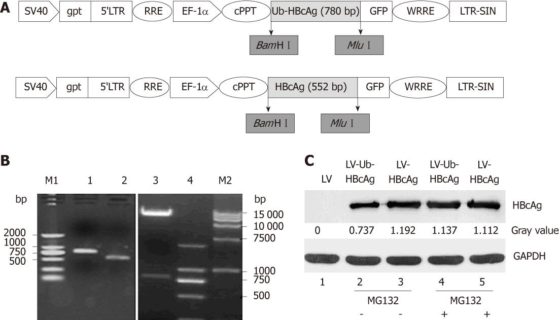
Figure 1 Schematic diagram, electrophoresis of ubiquitinated hepatitis B virus core antigen and HBcAg genes, pWPXLd-Ub-HBcAg digested by BamHIand MluI, and HBcAg protein expression (about 21 kDa).
A: Schematic diagram of pWPXLd vector; B: Lane 1, ubiquitinated hepatitis B virus core antigen (Ub-HBcAg) polymerase chain reaction (PCR) product (780 bp); lane 2, HBcAg PCR product (552 bp); lane 3, The digested products pWPXLd-Ub-HBcAg by BamHI and MluI; lane 4 and lane M1, DNA marker 2000; lane M2, DNA marker 15 000; C: 293T cells were transduced with lentiviral vector (LV), LV-Ub-HBcAg or LV-HBcAg and cultured for 48 h. MG-132 (10 mmol) was added for 24 h before harvesting the cells. Cell lysates (10 mg) were analyzed by immunoblotting with an anti-HBc antibody. Relative expression of HBcAg was calculated by a gray value.
- Citation: Chen JH, Yu YS, Chen XH, Liu HH, Zang GQ, Tang ZH. Enhancement of CTLs induced by DCs loaded with ubiquitinated hepatitis B virus core antigen. World J Gastroenterol 2012; 18(12): 1319-1327
- URL: https://www.wjgnet.com/1007-9327/full/v18/i12/1319.htm
- DOI: https://dx.doi.org/10.3748/wjg.v18.i12.1319


