Copyright
©2012 Baishideng Publishing Group Co.
World J Gastroenterol. Mar 28, 2012; 18(12): 1295-1307
Published online Mar 28, 2012. doi: 10.3748/wjg.v18.i12.1295
Published online Mar 28, 2012. doi: 10.3748/wjg.v18.i12.1295
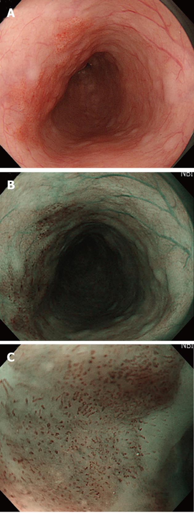
Figure 1 The carcinoma visualized in esophagus.
A: Carcinoma in esophagus is difficult to identify by conventional white light; B: Carcinoma in esophagus can be easily recognized by narrow-band imaging (NBI) as well-demarcated brownish area; C: Intraepithelial papillary capillary loop can be observed by magnifying endoscopy with NBI at the edge of the tumor.
- Citation: Chai NL, Ling-Hu EQ, Morita Y, Obata D, Toyonaga T, Azuma T, Wu BY. Magnifying endoscopy in upper gastroenterology for assessing lesions before completing endoscopic removal. World J Gastroenterol 2012; 18(12): 1295-1307
- URL: https://www.wjgnet.com/1007-9327/full/v18/i12/1295.htm
- DOI: https://dx.doi.org/10.3748/wjg.v18.i12.1295


