Copyright
©2012 Baishideng Publishing Group Co.
World J Gastroenterol. Mar 21, 2012; 18(11): 1273-1278
Published online Mar 21, 2012. doi: 10.3748/wjg.v18.i11.1273
Published online Mar 21, 2012. doi: 10.3748/wjg.v18.i11.1273
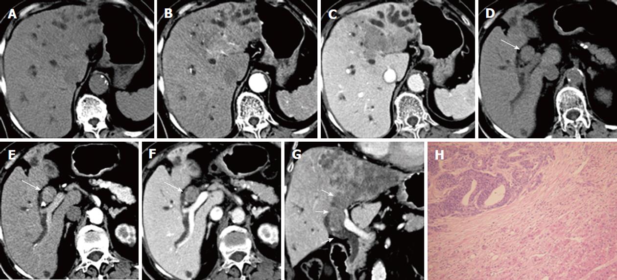
Figure 2 A 75-year-old woman with liver metastasis from colon cancer.
A lobulated, ill-defined hypodense tumor in the left hepatic lobe is noted on pre-contrast computed tomography (CT) scans (A), which shows early enhancement in the hepatic artery phase (B) and a low density in the portal phase (C). The bile duct tumor thrombus (BDTT) (arrow) shows similar enhancement patterns with the intrahepatic tumor on precontrast CT scan (D), hepatic artery phase (E) and portal phase scan (F). The coronal reconstruction image in the portal phase shows that BDTT (arrows) is contiguous with the intrahepatic tumor (G). Intraductal tumor thrombus is proved to be metastatic adenocarcinoma pathologically (H), hematoxylin and eosin stain, × 100.
- Citation: Liu QY, Lin XF, Li HG, Gao M, Zhang WD. Tumors with macroscopic bile duct thrombi in non-HCC patients: Dynamic multi-phase MSCT findings. World J Gastroenterol 2012; 18(11): 1273-1278
- URL: https://www.wjgnet.com/1007-9327/full/v18/i11/1273.htm
- DOI: https://dx.doi.org/10.3748/wjg.v18.i11.1273


