Copyright
©2012 Baishideng Publishing Group Co.
World J Gastroenterol. Jan 7, 2012; 18(1): 1-15
Published online Jan 7, 2012. doi: 10.3748/wjg.v18.i1.1
Published online Jan 7, 2012. doi: 10.3748/wjg.v18.i1.1
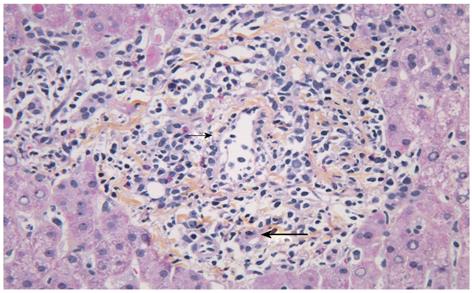
Figure 3 Histological changes seen in a biopsy with acute cellular rejection in a patient with primary sclerosing cholangitis.
Portal tract with mixed inflammatory infiltrate containing blastic lymphocytes and eosinophils. Subendothelial localization of the inflammatory cells in a portal vein branch (small arrow). Inflammation of small bile duct (large arrow). (Original magnification, x 400).
- Citation: Fosby B, Karlsen TH, Melum E. Recurrence and rejection in liver transplantation for primary sclerosing cholangitis. World J Gastroenterol 2012; 18(1): 1-15
- URL: https://www.wjgnet.com/1007-9327/full/v18/i1/1.htm
- DOI: https://dx.doi.org/10.3748/wjg.v18.i1.1


