Copyright
©2011 Baishideng Publishing Group Co.
World J Gastroenterol. Mar 7, 2011; 17(9): 1234-1236
Published online Mar 7, 2011. doi: 10.3748/wjg.v17.i9.1234
Published online Mar 7, 2011. doi: 10.3748/wjg.v17.i9.1234
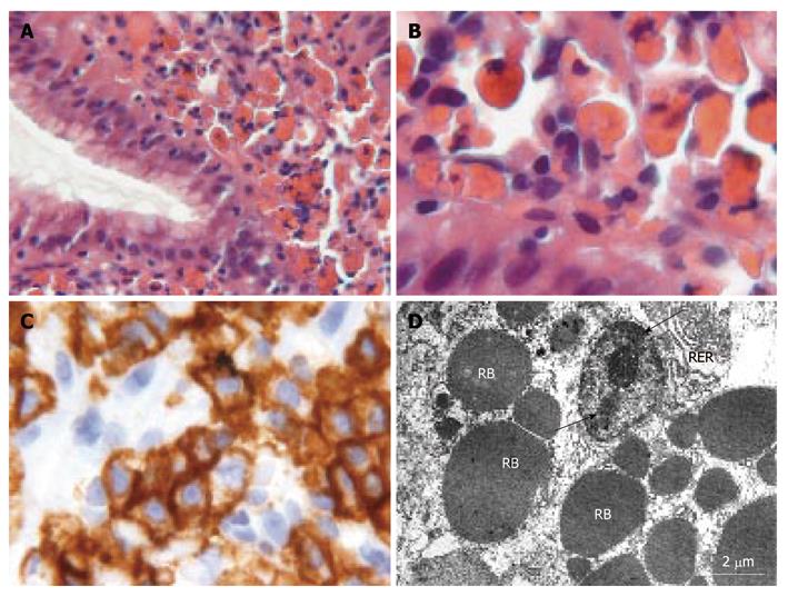
Figure 1 Histological, immunohistochemical and ultrastructural features of Russell body gastritis.
A: Gastroesophageal junction sample showing monomorphous cells with eosinophilic inclusions: Russell bodies; B: Higher magnification of plasma cells with crystalline inclusions; C: Immunohistochemical reactivity of plasma cells for CD138 antibody; D: Electron micrograph of a plasma cell with typical condensation pattern of nuclear chromatin (arrows) and well-developed rough endoplasmic reticulum (RER). Several Russell bodies (RB) with 1-5 μm diameter are present in dilated RER cisternae.
- Citation: Gobbo AD, Elli L, Braidotti P, Nuovo FD, Bosari S, Romagnoli S. Helicobacter pylori-negative Russell body gastritis: Case report. World J Gastroenterol 2011; 17(9): 1234-1236
- URL: https://www.wjgnet.com/1007-9327/full/v17/i9/1234.htm
- DOI: https://dx.doi.org/10.3748/wjg.v17.i9.1234


