Copyright
©2011 Baishideng Publishing Group Co.
World J Gastroenterol. Mar 7, 2011; 17(9): 1192-1198
Published online Mar 7, 2011. doi: 10.3748/wjg.v17.i9.1192
Published online Mar 7, 2011. doi: 10.3748/wjg.v17.i9.1192
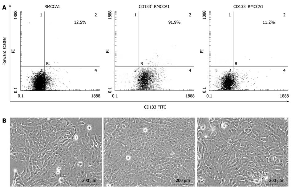
Figure 3 Isolation of CD133+ and CD133- cholangiocarcinoma cells.
A: The percentage of CD133-expressing cells in the original RMCCA1 cell populations, the flushed-out fractions (CD133+ RMCCA1 cells) and the flow-through (CD133- RMCCA1 cells) were analyzed by fluorescence-activated cell sorting; B: The morphology of original, CD133+ and CD133- RMCCA1 cells is demonstrated under a phase contrast microscope at 200 × magnification. FITC: Fluorescein isothiocyanate.
- Citation: Leelawat K, Thongtawee T, Narong S, Subwongcharoen S, Treepongkaruna SA. Strong expression of CD133 is associated with increased cholangiocarcinoma progression. World J Gastroenterol 2011; 17(9): 1192-1198
- URL: https://www.wjgnet.com/1007-9327/full/v17/i9/1192.htm
- DOI: https://dx.doi.org/10.3748/wjg.v17.i9.1192


