Copyright
©2011 Baishideng Publishing Group Co.
World J Gastroenterol. Feb 28, 2011; 17(8): 968-975
Published online Feb 28, 2011. doi: 10.3748/wjg.v17.i8.968
Published online Feb 28, 2011. doi: 10.3748/wjg.v17.i8.968
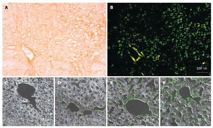
Figure 5 Histological characterization of amyloidosis mice.
Light microscopy of a liver section from a 6-mo-old K mouse stained with Congo red (A) and the same section under polarized light showing green birefringence (B); Confocal microscopy of liver sections with immunolocalization of mutant hapoA-II using an anti hapoA-II antibody and CY2 as the secondary antibody (green fluorescence): Control mouse, 8 mo old (C); K mouse, 2 mo old (D); K mouse, 6 mo old (E); F mouse, 8 mo old (F).
- Citation: Bastard C, Bosisio MR, Chabert M, Kalopissis AD, Mahrouf-Yorgov M, Gilgenkrantz H, Mueller S, Sandrin L. Transient micro-elastography: A novel non-invasive approach to measure liver stiffness in mice. World J Gastroenterol 2011; 17(8): 968-975
- URL: https://www.wjgnet.com/1007-9327/full/v17/i8/968.htm
- DOI: https://dx.doi.org/10.3748/wjg.v17.i8.968


