Copyright
©2011 Baishideng Publishing Group Co.
World J Gastroenterol. Feb 28, 2011; 17(8): 1051-1057
Published online Feb 28, 2011. doi: 10.3748/wjg.v17.i8.1051
Published online Feb 28, 2011. doi: 10.3748/wjg.v17.i8.1051
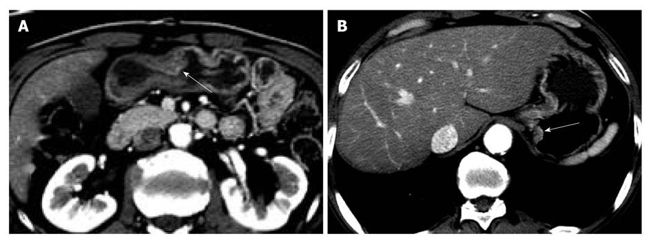
Figure 3 Early gastric cancer in a 62-year-old man.
The size of tumor was 2.2 cm at the longest diameter and the tumor extended to submucosal layer. A: The transverse computed tomography scan shows thickened wall with enhancement in greater curvature of gastric antrum (arrow), which was true lesion based on gastroscopy and pathologic examination; B: Reviewer 1 indicated a lesion (arrow) as a cancer focus because thickened wall was suspicious on blinded analysis. However, on unblinded analysis, focal lesion (arrow) in greater curvature of gastric antrum in lower gastric 1/3 segment was indicated correctly.
- Citation: Park KJ, Lee MW, Koo JH, Park Y, Kim H, Choi D, Lee SJ. Detection of early gastric cancer using hydro-stomach CT: Blinded vs unblinded analysis. World J Gastroenterol 2011; 17(8): 1051-1057
- URL: https://www.wjgnet.com/1007-9327/full/v17/i8/1051.htm
- DOI: https://dx.doi.org/10.3748/wjg.v17.i8.1051


