Copyright
©2011 Baishideng Publishing Group Co.
World J Gastroenterol. Feb 21, 2011; 17(7): 946-952
Published online Feb 21, 2011. doi: 10.3748/wjg.v17.i7.946
Published online Feb 21, 2011. doi: 10.3748/wjg.v17.i7.946
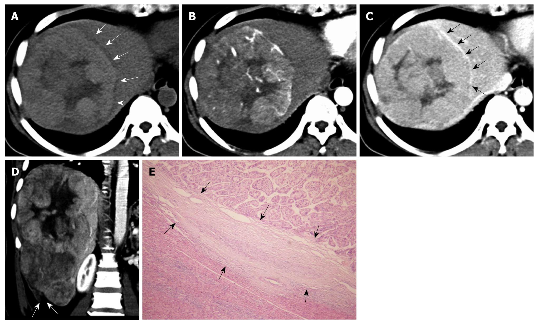
Figure 1 Primary clear cell carcinoma of the liver in a 47-year-old woman.
A: On pre-contrast computed tomography scan, the mass shows slight hyper-attenuation with a hypo-attenuation halo (arrows); B: At hepatic arterial phase, the mass shows early enhancement; C: At the equilibrium phase, the mass presents hypo-attenuation with rim enhancement (arrows); D: At portal venous phase, the reconstructed coronal image shows the mass with a discontinuous liver capsule (arrows) at Segment VI, indicating tumor rupture, which was surgically confirmed; E: Pathologically, the mass shows a pseudocapsule (arrows) (HE, × 100).
- Citation: Liu QY, Li HG, Gao M, Lin XF, Li Y, Chen JY. Primary clear cell carcinoma in the liver: CT and MRI findings. World J Gastroenterol 2011; 17(7): 946-952
- URL: https://www.wjgnet.com/1007-9327/full/v17/i7/946.htm
- DOI: https://dx.doi.org/10.3748/wjg.v17.i7.946


