Copyright
©2011 Baishideng Publishing Group Co.
World J Gastroenterol. Feb 14, 2011; 17(6): 789-795
Published online Feb 14, 2011. doi: 10.3748/wjg.v17.i6.789
Published online Feb 14, 2011. doi: 10.3748/wjg.v17.i6.789
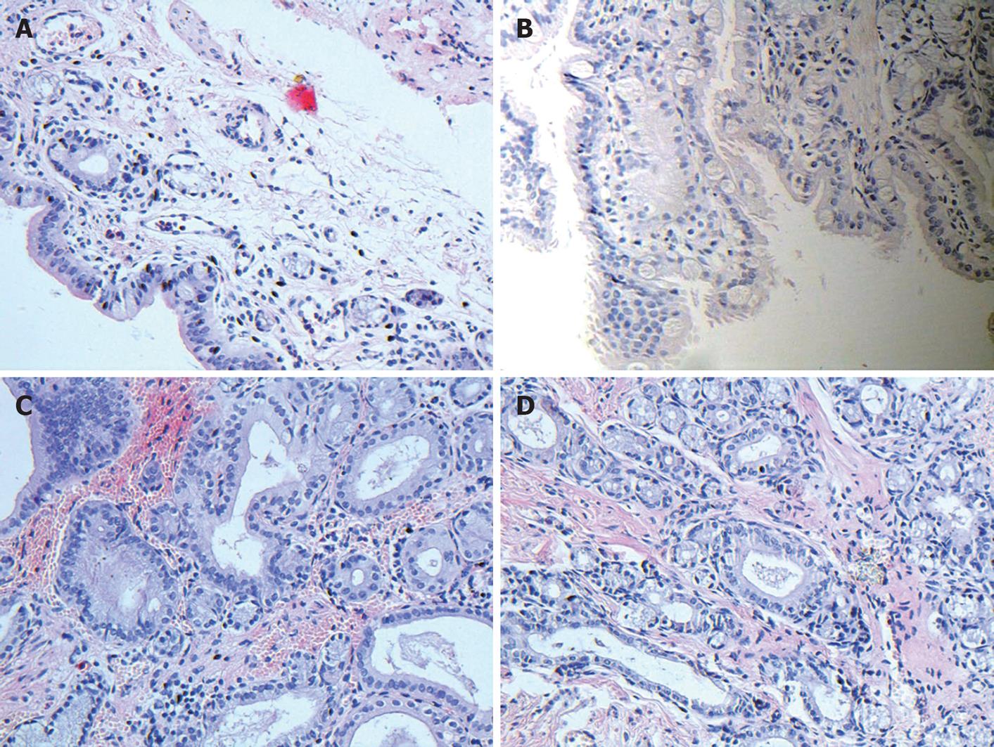
Figure 3 Histological examination of bile duct in all groups (original magnification, × 200).
A: Normal bile duct intima; B: Gland proliferation and infiltration of inflammatory cells in 2-mo group; C: Fibrous thickening of bile duct and dense infiltration of inflammatory cells in 3-mo group; D: Epithelial proliferation as well as intra- and extramural glandular hyperplasia in bile duct.
- Citation: Zhang XQ, Tian YH, Xu Z, Wang LX, Hou CS, Ling XF, Zhou XS. An end-to-end anastomosis model of guinea pig bile duct: A 6-mo observation. World J Gastroenterol 2011; 17(6): 789-795
- URL: https://www.wjgnet.com/1007-9327/full/v17/i6/789.htm
- DOI: https://dx.doi.org/10.3748/wjg.v17.i6.789


