Copyright
©2011 Baishideng Publishing Group Co.
World J Gastroenterol. Feb 7, 2011; 17(5): 677-680
Published online Feb 7, 2011. doi: 10.3748/wjg.v17.i5.677
Published online Feb 7, 2011. doi: 10.3748/wjg.v17.i5.677
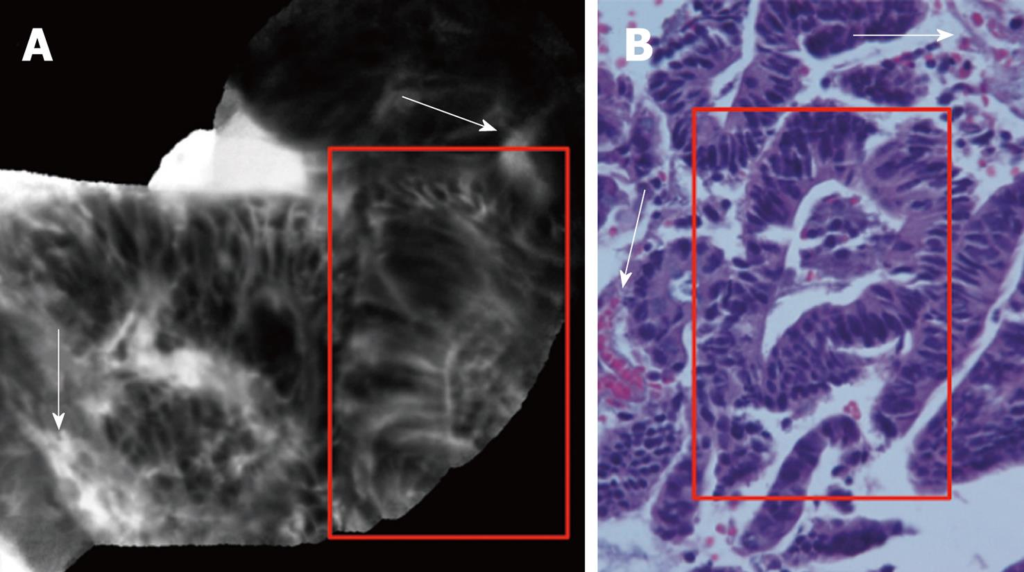
Figure 4 Confocal (A) and histological (B) images of dysplasia-associated lesional mass showing “dark” cells, with mucin depletion and goblet cell/crypt density attenuation; the architectural pattern is irregular, as well as the epithelial thickness, with villiform structures and “dark” epithelial border (red rectangles).
There is gross distortion of the vascular architecture with tortuous and dilated vessels (white arrows). The hematoxylin and eosin stain histology shows a low grade dysplasia (red rectangle; hematoxylin and eosin staining; original magnification, × 200).
- Citation: Palma GDD, Staibano S, Siciliano S, Maione F, Siano M, Esposito D, Persico G. In vivo characterization of DALM in ulcerative colitis with high-resolution probe-based confocal laser endomicroscopy. World J Gastroenterol 2011; 17(5): 677-680
- URL: https://www.wjgnet.com/1007-9327/full/v17/i5/677.htm
- DOI: https://dx.doi.org/10.3748/wjg.v17.i5.677


