Copyright
©2011 Baishideng Publishing Group Co.
World J Gastroenterol. Feb 7, 2011; 17(5): 677-680
Published online Feb 7, 2011. doi: 10.3748/wjg.v17.i5.677
Published online Feb 7, 2011. doi: 10.3748/wjg.v17.i5.677
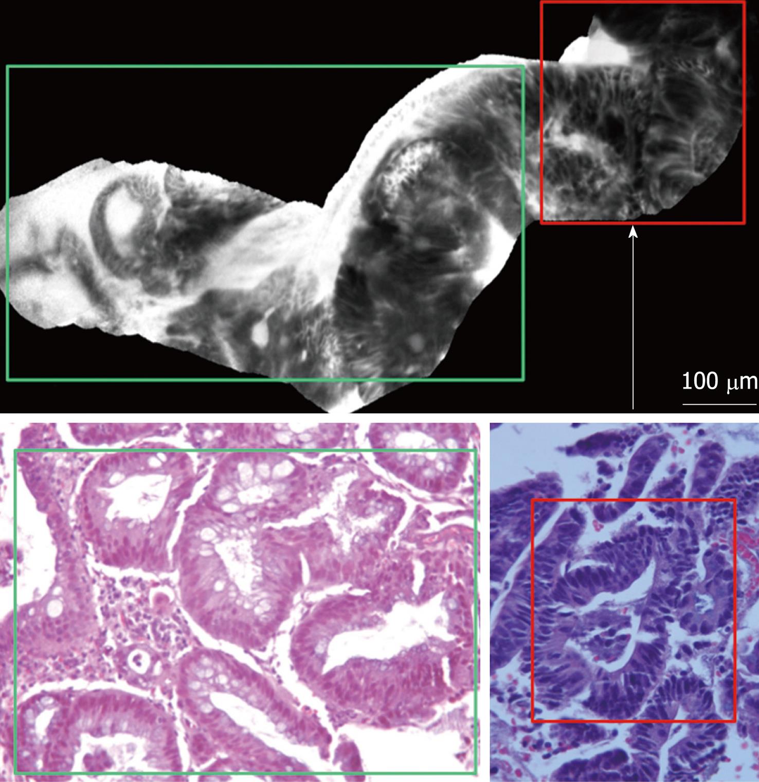
Figure 3 Confocal images of colonic mucosa evidencing the switch from the inflamed mucosa, to the neoplastic mucosa.
Inflamed mucosa (green rectangle) is characterized by dilation of crypt openings, enlarged spaces between crypt, and microvascular alterations with fluorescein leaks into the crypt lumen (white arrow) therefore making the lumen brighter than the surrounding epithelium. Dysplastic mucosa (red rectangle) is characterized by “dark” cells, irregular architectural patterns with villiform structures and a “dark” epithelial border. Histology images show high-power hematoxylin and eosin stain of the tissue sampled, evidencing respectively inflamed area with features suggestive of chronic ulcerative colitis (green rectangle) and low grade dysplasia (red rectangle). Magnification, × 200.
- Citation: Palma GDD, Staibano S, Siciliano S, Maione F, Siano M, Esposito D, Persico G. In vivo characterization of DALM in ulcerative colitis with high-resolution probe-based confocal laser endomicroscopy. World J Gastroenterol 2011; 17(5): 677-680
- URL: https://www.wjgnet.com/1007-9327/full/v17/i5/677.htm
- DOI: https://dx.doi.org/10.3748/wjg.v17.i5.677


