Copyright
©2011 Baishideng Publishing Group Co.
World J Gastroenterol. Feb 7, 2011; 17(5): 677-680
Published online Feb 7, 2011. doi: 10.3748/wjg.v17.i5.677
Published online Feb 7, 2011. doi: 10.3748/wjg.v17.i5.677
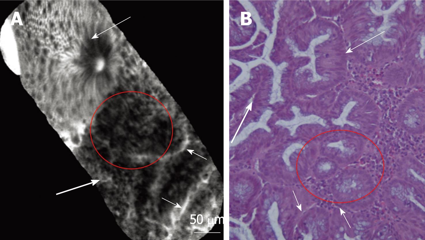
Figure 2 Confocal (A) and histological (B) images of colonic mucosa showing the switch from normal mucosa to inflamed mucosa.
Normal crypt architecture is classically represented by ordered and regular crypt orifices covered by a homogeneous epithelial layer with visible “black-hole” goblet cells within the subcellular matrix (long thin arrows). Inflamed mucosa showing irregular arrangement of crypts, crypt fusion (red circles) and capillaries alterations (short arrows) and inflammatory cells (lymphocytes: long thick arrows). Magnification, × 200.
- Citation: Palma GDD, Staibano S, Siciliano S, Maione F, Siano M, Esposito D, Persico G. In vivo characterization of DALM in ulcerative colitis with high-resolution probe-based confocal laser endomicroscopy. World J Gastroenterol 2011; 17(5): 677-680
- URL: https://www.wjgnet.com/1007-9327/full/v17/i5/677.htm
- DOI: https://dx.doi.org/10.3748/wjg.v17.i5.677


