Copyright
©2011 Baishideng Publishing Group Co.
World J Gastroenterol. Feb 7, 2011; 17(5): 677-680
Published online Feb 7, 2011. doi: 10.3748/wjg.v17.i5.677
Published online Feb 7, 2011. doi: 10.3748/wjg.v17.i5.677
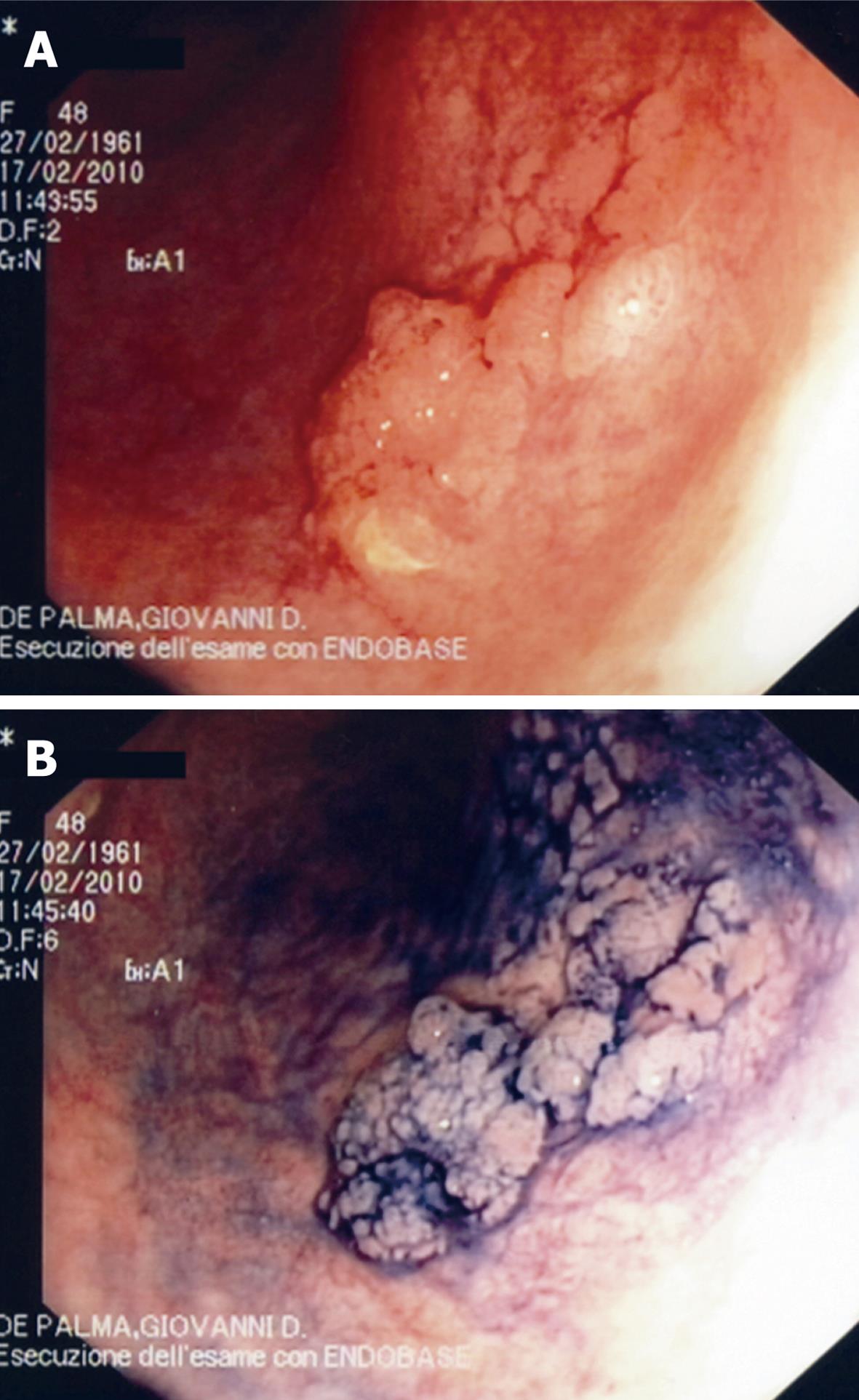
Figure 1 Conventional “white light” imaging of a plaque-like lesion of sigmoid colon in ulcerative colitis (A) and 0.
5% indigo carmine chromoscopy of the lesion (B). The lesion is “unmasked” and clearly delineated.
- Citation: Palma GDD, Staibano S, Siciliano S, Maione F, Siano M, Esposito D, Persico G. In vivo characterization of DALM in ulcerative colitis with high-resolution probe-based confocal laser endomicroscopy. World J Gastroenterol 2011; 17(5): 677-680
- URL: https://www.wjgnet.com/1007-9327/full/v17/i5/677.htm
- DOI: https://dx.doi.org/10.3748/wjg.v17.i5.677


