Copyright
©2011 Baishideng Publishing Group Co.
World J Gastroenterol. Dec 28, 2011; 17(48): 5305-5309
Published online Dec 28, 2011. doi: 10.3748/wjg.v17.i48.5305
Published online Dec 28, 2011. doi: 10.3748/wjg.v17.i48.5305
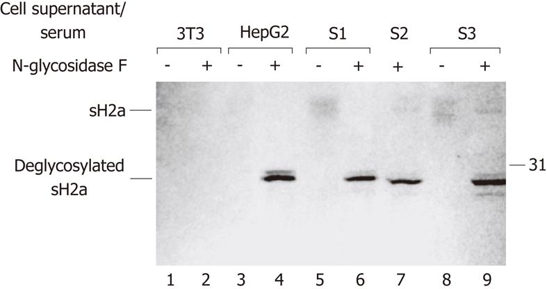
Figure 1 sH2a is detected in normal human sera.
Cell supernatants from 90 mm petri-dishes of NIH 3T3 (lanes 1-2) or HepG2 cells (lanes 3-4) or 0.3 mL of normal human sera from 3 donors (S1, lanes 5 and 6; S2, lane 7; S3, lanes 8 and 9) were immunoprecipitated with polyclonal anti-H2a carboxyterminal antibodies that were crosslinked to protein A-agarose, and the immunoprecipitates were subjected to 12% sodium dodecyl sulfate polyacrylamide gel electrophoresis. Immunoblotting was then done with anti-H2a carboxyterminal antibody followed by horseradish peroxidase-conjugated goat anti-rabbit IgG and color development with 3,3’,5,5’-tetramethylbenzidine. Samples on lanes 2, 4, 6, 7 and 9 were treated with N-glycosidase-F after immunoprecipitation. On the right is the molecular weight of a protein standard in kilodaltons. On the left are the migrations of sH2a before or after deglycosylation.
- Citation: Benyair R, Kondratyev M, Veselkin E, Tolchinsky S, Shenkman M, Lurie Y, Lederkremer GZ. Constant serum levels of secreted asialoglycoprotein receptor sH2a and decrease with cirrhosis. World J Gastroenterol 2011; 17(48): 5305-5309
- URL: https://www.wjgnet.com/1007-9327/full/v17/i48/5305.htm
- DOI: https://dx.doi.org/10.3748/wjg.v17.i48.5305


