Copyright
©2011 Baishideng Publishing Group Co.
World J Gastroenterol. Dec 21, 2011; 17(47): 5203-5213
Published online Dec 21, 2011. doi: 10.3748/wjg.v17.i47.5203
Published online Dec 21, 2011. doi: 10.3748/wjg.v17.i47.5203
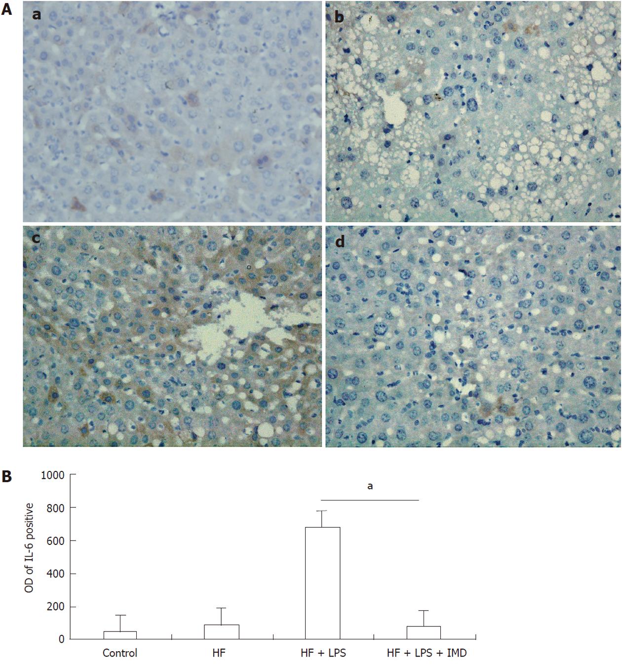
Figure 3 Interleukin-6 expression was assessed by immunohistochemistry.
A: Positive staining was observed in hepatocytes in the control group (a), HF group (b), LPS-induced HF group (c) and IMD-treated group (d). B: The optical density (OD) of interleukin-6 (IL-6)-positive areas was measured with ImageProplu6.0 (aP < 0.05). HF: High-fat; LPS: Lipopolysaccharide; IMD: IKK2 inhibitor.
- Citation: Wei J, Shi M, Wu WQ, Xu H, Wang T, Wang N, Ma JL, Wang YG. IκB kinase-beta inhibitor attenuates hepatic fibrosis in mice. World J Gastroenterol 2011; 17(47): 5203-5213
- URL: https://www.wjgnet.com/1007-9327/full/v17/i47/5203.htm
- DOI: https://dx.doi.org/10.3748/wjg.v17.i47.5203


