Copyright
©2011 Baishideng Publishing Group Co.
World J Gastroenterol. Dec 14, 2011; 17(46): 5110-5116
Published online Dec 14, 2011. doi: 10.3748/wjg.v17.i46.5110
Published online Dec 14, 2011. doi: 10.3748/wjg.v17.i46.5110
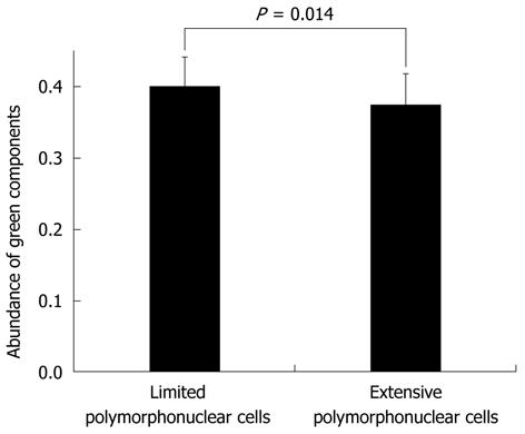
Figure 6 Comparison of limited (combined grades 0 and 1) and extensive (combined grades 2 and 3) polymorphonuclear cell infiltration of the green color components in Mayo endoscopic subscore-0.
The mean number of green color components for images classified into the amount of polymorphonuclear cell infiltration in Mayo endoscopic subscore (MES)-0 is 0.399 ± 0.042 for limited and 0.375 ± 0.044 for extensive infiltration. There are significant differences in green color component values between limited and extensive polymorphonuclear cell infiltration in MES-0 (P = 0.014, by Student’s t-test).
- Citation: Osada T, Arakawa A, Sakamoto N, Ueyama H, Shibuya T, Ogihara T, Yao T, Watanabe S. Autofluorescence imaging endoscopy for identification and assessment of inflammatory ulcerative colitis. World J Gastroenterol 2011; 17(46): 5110-5116
- URL: https://www.wjgnet.com/1007-9327/full/v17/i46/5110.htm
- DOI: https://dx.doi.org/10.3748/wjg.v17.i46.5110


