Copyright
©2011 Baishideng Publishing Group Co.
World J Gastroenterol. Dec 14, 2011; 17(46): 5110-5116
Published online Dec 14, 2011. doi: 10.3748/wjg.v17.i46.5110
Published online Dec 14, 2011. doi: 10.3748/wjg.v17.i46.5110
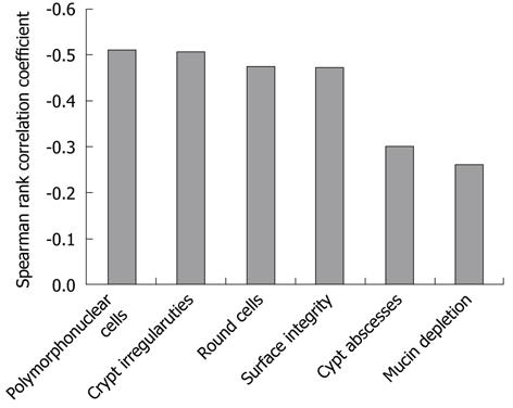
Figure 5 Comparison of histology scores, six histological characteristic ulcerative colitis features and the green color component on autofluorescence imaging.
The autofluorescence imaging green color component is relatively well correlated with the scores for polymorphonuclear cells in the lamina propria (r = -0.51, P < 0.01) and crypt architectural irregularities (r = -0.51, P < 0.01), but not with the scores for crypt abscesses (r = -0.30, P < 0.01), and mucin depletion (r = -0.26, P < 0.01) based on Spearman rank correlation coefficient.
- Citation: Osada T, Arakawa A, Sakamoto N, Ueyama H, Shibuya T, Ogihara T, Yao T, Watanabe S. Autofluorescence imaging endoscopy for identification and assessment of inflammatory ulcerative colitis. World J Gastroenterol 2011; 17(46): 5110-5116
- URL: https://www.wjgnet.com/1007-9327/full/v17/i46/5110.htm
- DOI: https://dx.doi.org/10.3748/wjg.v17.i46.5110


