Copyright
©2011 Baishideng Publishing Group Co.
World J Gastroenterol. Dec 14, 2011; 17(46): 5110-5116
Published online Dec 14, 2011. doi: 10.3748/wjg.v17.i46.5110
Published online Dec 14, 2011. doi: 10.3748/wjg.v17.i46.5110
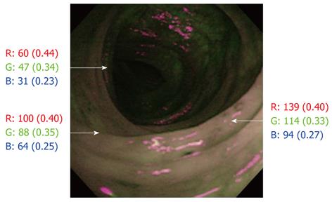
Figure 1 Endoscopic photograph by autofluorescence imaging showing the actual counts of red, green and blue colors based on the RGB additive model in different mucosal areas.
The actual counts of red, green and blue colors varied depending on the brightness of the examined area, but the abundance of the actual counts, in parentheses, was approximately constant in the same picture.
- Citation: Osada T, Arakawa A, Sakamoto N, Ueyama H, Shibuya T, Ogihara T, Yao T, Watanabe S. Autofluorescence imaging endoscopy for identification and assessment of inflammatory ulcerative colitis. World J Gastroenterol 2011; 17(46): 5110-5116
- URL: https://www.wjgnet.com/1007-9327/full/v17/i46/5110.htm
- DOI: https://dx.doi.org/10.3748/wjg.v17.i46.5110


