Copyright
©2011 Baishideng Publishing Group Co.
World J Gastroenterol. Dec 7, 2011; 17(45): 4999-5006
Published online Dec 7, 2011. doi: 10.3748/wjg.v17.i45.4999
Published online Dec 7, 2011. doi: 10.3748/wjg.v17.i45.4999
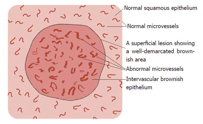
Figure 1 Schema of the magnified endoscopic features of a superficial lesion and its surrounding mucosa as detected by narrow-band imaging.
The superficial lesion shows an intervascular brownish epithelium and abnormal microvessels exhibiting dilation, proliferation and irregularities.
- Citation: Yoshimura N, Goda K, Tajiri H, Yoshida Y, Kato T, Seino Y, Ikegami M, Urashima M. Diagnostic utility of narrow-band imaging endoscopy for pharyngeal superficial carcinoma. World J Gastroenterol 2011; 17(45): 4999-5006
- URL: https://www.wjgnet.com/1007-9327/full/v17/i45/4999.htm
- DOI: https://dx.doi.org/10.3748/wjg.v17.i45.4999


