Copyright
©2011 Baishideng Publishing Group Co.
World J Gastroenterol. Dec 7, 2011; 17(45): 4952-4959
Published online Dec 7, 2011. doi: 10.3748/wjg.v17.i45.4952
Published online Dec 7, 2011. doi: 10.3748/wjg.v17.i45.4952
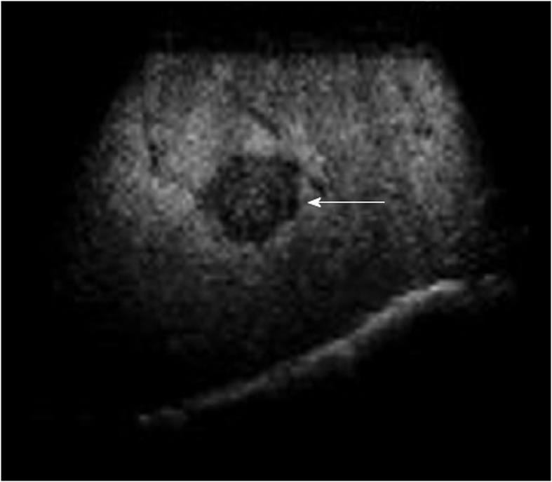
Figure 3 A 70-year-old man with a 2.
0-cm hepatocellular carcinoma nodule located in segment 6 of the liver. Contrast-enhanced ultrasound using sonazoid shows the defect (arrow) imaging in post-vascular phase. The defect lesion can be targeted for insertion of a single needle by extending the time limitation.
- Citation: Minami Y, Kudo M. Review of dynamic contrast-enhanced ultrasound guidance in ablation therapy for hepatocellular carcinoma. World J Gastroenterol 2011; 17(45): 4952-4959
- URL: https://www.wjgnet.com/1007-9327/full/v17/i45/4952.htm
- DOI: https://dx.doi.org/10.3748/wjg.v17.i45.4952


