Copyright
©2011 Baishideng Publishing Group Co.
World J Gastroenterol. Nov 28, 2011; 17(44): 4875-4882
Published online Nov 28, 2011. doi: 10.3748/wjg.v17.i44.4875
Published online Nov 28, 2011. doi: 10.3748/wjg.v17.i44.4875
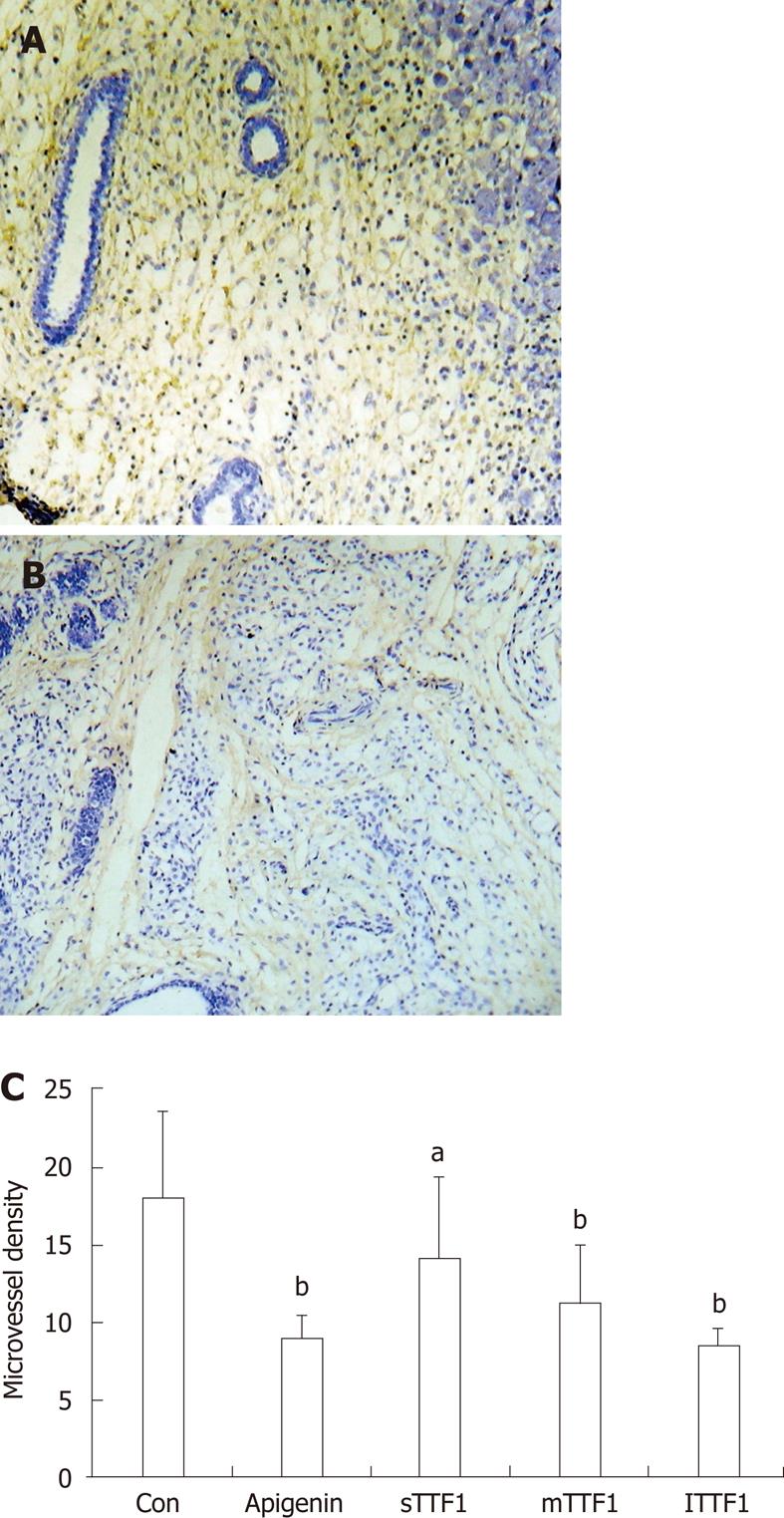
Figure 4 Microvessels in tumor tissue angiogenesis in HepG-2-transplanted nude mice.
Brown staining indicates CD34 positive cells. A: Control, HepG-2-transplanted nude mouse treated with normal saline; B: TTF1, HepG-2-transplanted nude mouse treated with TTF1; C: Different doses of compound were used as treatments in the HepG-2-transplanted nude mice as follows: sTTF1 (5 μmol/kg); mTTF1 (10 μmol/kg); lTTF1 (20 μmol/kg); apigenin (40 μmol/kg); and Con (control group treated with normal saline). aP < 0.05, bP < 0.01 vs control group.
- Citation: Liu C, Li XW, Cui LM, Li LC, Chen LY, Zhang XW. Inhibition of tumor angiogenesis by TTF1 from extract of herbal medicine. World J Gastroenterol 2011; 17(44): 4875-4882
- URL: https://www.wjgnet.com/1007-9327/full/v17/i44/4875.htm
- DOI: https://dx.doi.org/10.3748/wjg.v17.i44.4875


