Copyright
©2011 Baishideng Publishing Group Co.
World J Gastroenterol. Nov 21, 2011; 17(43): 4757-4771
Published online Nov 21, 2011. doi: 10.3748/wjg.v17.i43.4757
Published online Nov 21, 2011. doi: 10.3748/wjg.v17.i43.4757
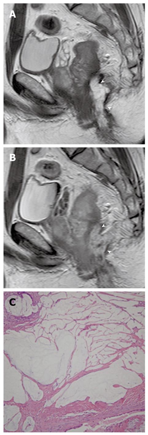
Figure 16 Perianal mucinous adenocarcinoma in a 41-year-old man.
A: Sagittal T2-weighted MR image shows a hyperintense mass (arrowheads) in the perianal area; B: Sagittal contrast-enhanced T1-weighted MR image shows mesh-like internal enhancement (arrowheads) within the mass; C: Photomicrograph image (original magnification, × 100; HE stain) shows large pools of extracellular mucin and tumor cells. MR: Magnetic resonance; HE: Hematoxylin and eosin.
- Citation: Lee NK, Kim S, Kim HS, Jeon TY, Kim GH, Kim DU, Park DY, Kim TU, Kang DH. Spectrum of mucin-producing neoplastic conditions of the abdomen and pelvis: Cross-sectional imaging evaluation. World J Gastroenterol 2011; 17(43): 4757-4771
- URL: https://www.wjgnet.com/1007-9327/full/v17/i43/4757.htm
- DOI: https://dx.doi.org/10.3748/wjg.v17.i43.4757


