Copyright
©2011 Baishideng Publishing Group Co.
World J Gastroenterol. Nov 21, 2011; 17(43): 4757-4771
Published online Nov 21, 2011. doi: 10.3748/wjg.v17.i43.4757
Published online Nov 21, 2011. doi: 10.3748/wjg.v17.i43.4757
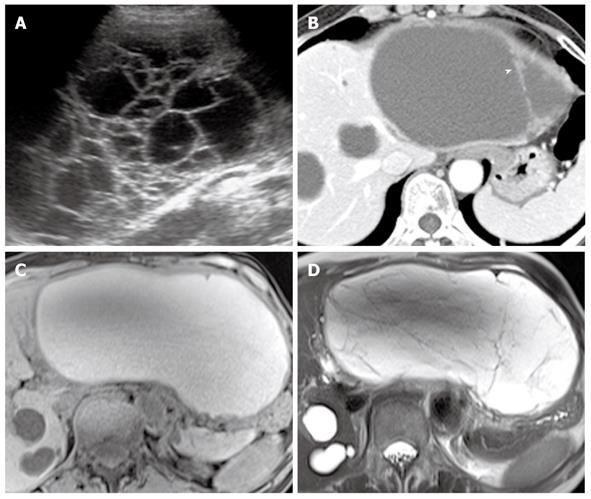
Figure 3 Biliary cystadenoma in the liver of a 55-year-old woman.
A: Ultrasound image shows a multiseptated cystic mass in the liver; B: Contrast-enhanced CT image shows a well-circumscribed cystic liver mass, which has similar attenuation to that of hepatic cysts. Note the internal septa with nodular thickening (arrowhead); C, D: T1- (C) and T2-weighted (D) MR images show a multiseptated cystic mass with hyperintensity. CT: Computed tomography; MR: Magnetic resonance.
- Citation: Lee NK, Kim S, Kim HS, Jeon TY, Kim GH, Kim DU, Park DY, Kim TU, Kang DH. Spectrum of mucin-producing neoplastic conditions of the abdomen and pelvis: Cross-sectional imaging evaluation. World J Gastroenterol 2011; 17(43): 4757-4771
- URL: https://www.wjgnet.com/1007-9327/full/v17/i43/4757.htm
- DOI: https://dx.doi.org/10.3748/wjg.v17.i43.4757


