Copyright
©2011 Baishideng Publishing Group Co.
World J Gastroenterol. Nov 14, 2011; 17(42): 4734-4738
Published online Nov 14, 2011. doi: 10.3748/wjg.v17.i42.4734
Published online Nov 14, 2011. doi: 10.3748/wjg.v17.i42.4734
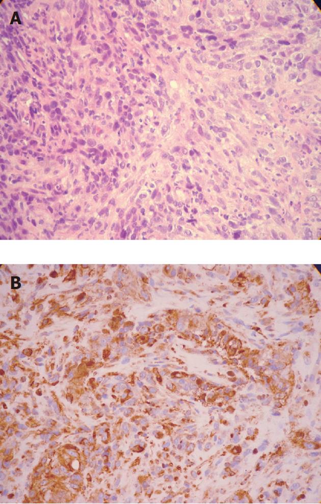
Figure 3 Histopathological findings.
A: Hematoxylin and eosin staining discloses crowded rounded or elongated cells within distinct cytoplasm and hyperchromatic nuclei (magnification, x 100); B: Immunohistochemical study for HMB45 (anti-melanoma protein mAb). The positive reaction (brown cytoplasmic reaction) supports the diagnosis of malignant melanoma (magnification, x 100).
- Citation: Machado J, Ministro P, Araújo R, Cancela E, Castanheira A, Silva A. Primary malignant melanoma of the esophagus: A case report. World J Gastroenterol 2011; 17(42): 4734-4738
- URL: https://www.wjgnet.com/1007-9327/full/v17/i42/4734.htm
- DOI: https://dx.doi.org/10.3748/wjg.v17.i42.4734


