Copyright
©2011 Baishideng Publishing Group Co.
World J Gastroenterol. Nov 14, 2011; 17(42): 4729-4733
Published online Nov 14, 2011. doi: 10.3748/wjg.v17.i42.4729
Published online Nov 14, 2011. doi: 10.3748/wjg.v17.i42.4729
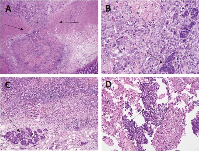
Figure 1 Hematoxylin and eosin-stained sections of tumor.
A: The tumor (asterisk) is submucosal with extension through inner and outer muscularis propria (arrows), 100 ×; B: Intimate mixing of squamous (asterisk) and neuroendocrine (arrow) components, 400 ×; C: Lymph node metastasis of both components (squamous, asterisk; neuroendocrine, arrow), 100 ×; D: Liver fine needle aspirate showing predominantly neuroendocrine cells (arrow) with background bland hepatocytes, 200 ×.
- Citation: Wentz SC, Vnencak-Jones C, Chopp WV. Neuroendocrine and squamous colonic composite carcinoma: Case report with molecular analysis. World J Gastroenterol 2011; 17(42): 4729-4733
- URL: https://www.wjgnet.com/1007-9327/full/v17/i42/4729.htm
- DOI: https://dx.doi.org/10.3748/wjg.v17.i42.4729


