Copyright
©2011 Baishideng Publishing Group Co.
World J Gastroenterol. Oct 28, 2011; 17(40): 4479-4487
Published online Oct 28, 2011. doi: 10.3748/wjg.v17.i40.4479
Published online Oct 28, 2011. doi: 10.3748/wjg.v17.i40.4479
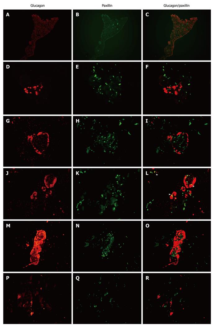
Figure 4 Immunofluorescent localization of paxillin and glucagon in the pancreas of E15.
5, E18.5, P0, P7, P21 and adult rats. The paxillin antibody was detected with an fluorescein isothiocyanate (green)-labeled secondary antibody and the glucagon antibody was detected with a Cy3 (red)-labeled secondary antibody. Overlap between paxillin (green) and glucagon (red) labeling is indicated by arrowheads. Original magnification 400 ×. A-C: E15.5; D-F: E18.5; G-I: P0; J-L: P7; M-O: P21; P-R: Adult.
- Citation: Guo J, Liu LJ, Yuan L, Wang N, De W. Expression and localization of paxillin in rat pancreas during development. World J Gastroenterol 2011; 17(40): 4479-4487
- URL: https://www.wjgnet.com/1007-9327/full/v17/i40/4479.htm
- DOI: https://dx.doi.org/10.3748/wjg.v17.i40.4479


