Copyright
©2011 Baishideng Publishing Group Co.
World J Gastroenterol. Oct 28, 2011; 17(40): 4479-4487
Published online Oct 28, 2011. doi: 10.3748/wjg.v17.i40.4479
Published online Oct 28, 2011. doi: 10.3748/wjg.v17.i40.4479
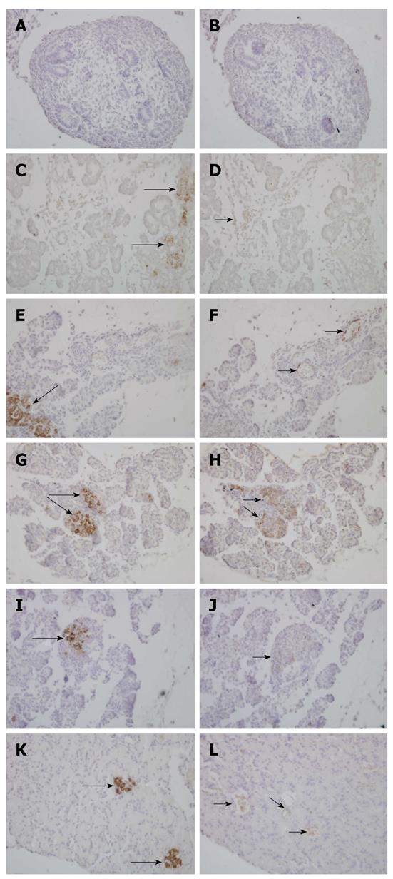
Figure 2 Immunohistochemical analysis of insulin and paxillin in the serial sections of the rat pancreas at E15.
5 (A, B), E18.5 (C, D), P0 (E, F), P14 (G, H), P21 (I, J) and adult (K, L). Adjacent pancreatic sections from six developmental stages were stained with antibodies against insulin (left lane) and paxillin (right lane), respectively. We acquired images using an OLYMPUS DP70 digital camera. Strong cytoplasmic staining was observed for insulin (long arrows) for five stages except E15.5. Immunolocalization for the paxillin revealed a sporadic positive staining (short arrows) in the pancreas. In E18.5 and P0 rats, some cells in the pancreas, but not in islets, were stained. As shown in G-J, at P14 and P21 paxillin was mainly localized in islets. All magnifications are × 400.
- Citation: Guo J, Liu LJ, Yuan L, Wang N, De W. Expression and localization of paxillin in rat pancreas during development. World J Gastroenterol 2011; 17(40): 4479-4487
- URL: https://www.wjgnet.com/1007-9327/full/v17/i40/4479.htm
- DOI: https://dx.doi.org/10.3748/wjg.v17.i40.4479


