Copyright
©2011 Baishideng Publishing Group Co.
World J Gastroenterol. Oct 28, 2011; 17(40): 4470-4478
Published online Oct 28, 2011. doi: 10.3748/wjg.v17.i40.4470
Published online Oct 28, 2011. doi: 10.3748/wjg.v17.i40.4470
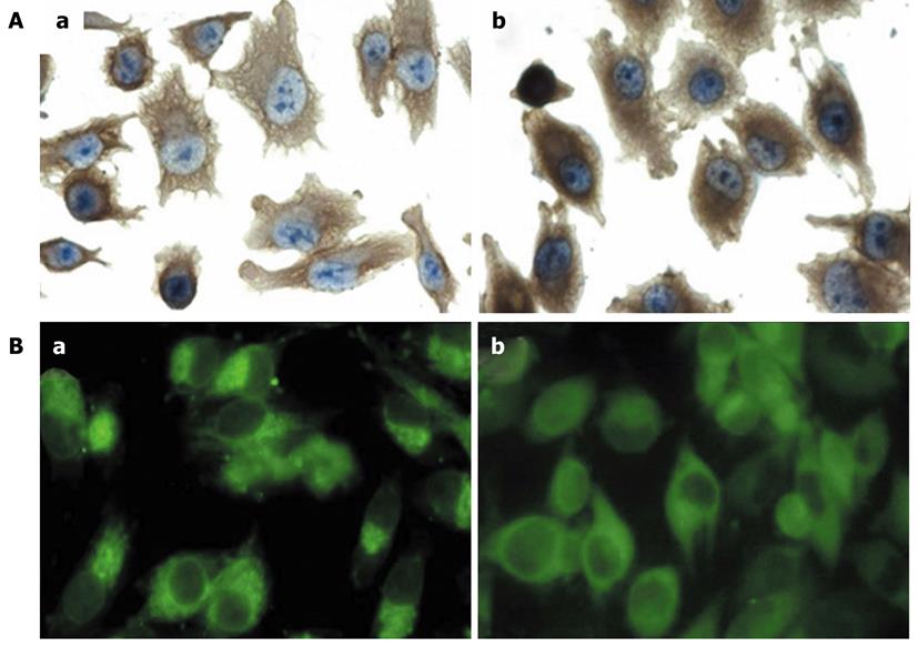
Figure 2 Fascin overexpression induces alteration of cell morphology and cytoskeleton.
A: Immunohistochemical analysis of actin distribution in fascin-overexpressing cells and vector control cells (× 400). (a) MIA PaCa-2 Fascin cells were more polarized with elongated membrane projections. Actin filaments were distributed as bundles in the cytoplasm which protruded into membrane projections in MIA PaCa-2 Fascin cells. (b) MIA PaCa-2 Vector cells showed a diffuse actin distribution; B: Immunofluorescence analysis of actin distribution in fascin-overexpressing cells and vector control cells (× 400). (a) Actin accumulated in a polarized manner in MIA PaCa-2 Fascin cells, whereas (b) MIA PaCa-2 Vector cells demonstrated a diffuse actin distribution.
- Citation: Xu YF, Yu SN, Lu ZH, Liu JP, Chen J. Fascin promotes the motility and invasiveness of pancreatic cancer cells. World J Gastroenterol 2011; 17(40): 4470-4478
- URL: https://www.wjgnet.com/1007-9327/full/v17/i40/4470.htm
- DOI: https://dx.doi.org/10.3748/wjg.v17.i40.4470


