Copyright
©2011 Baishideng Publishing Group Co.
World J Gastroenterol. Oct 28, 2011; 17(40): 4456-4460
Published online Oct 28, 2011. doi: 10.3748/wjg.v17.i40.4456
Published online Oct 28, 2011. doi: 10.3748/wjg.v17.i40.4456
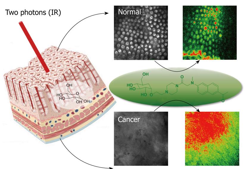
Figure 2 Images of normal tissue (above) and cancer tissue (below) treated with AG2.
Normal tissues were incubated in artificial cerebrospinal fluid (ACSF) for 4 h, and cancer tissues were incubated in ACSF for 4 h, after which AG2 uptake was monitored. Right-side images are bright-field images, left-side images are pseudocolored two-photon microscopy (TPM) images obtained after incubation with AG2 for 4 h. The TPM images were obtained at a depth of 100 μm by collecting the two-photon excited fluorescence spectra in the range of 520-620 nm on excitation with fs pulses at 780 nm. IR: Infrared.
- Citation: Cho HJ, Chun HJ, Kim ES, Cho BR. Multiphoton microscopy: An introduction to gastroenterologists. World J Gastroenterol 2011; 17(40): 4456-4460
- URL: https://www.wjgnet.com/1007-9327/full/v17/i40/4456.htm
- DOI: https://dx.doi.org/10.3748/wjg.v17.i40.4456


