Copyright
©2011 Baishideng Publishing Group Co.
World J Gastroenterol. Oct 28, 2011; 17(40): 4456-4460
Published online Oct 28, 2011. doi: 10.3748/wjg.v17.i40.4456
Published online Oct 28, 2011. doi: 10.3748/wjg.v17.i40.4456
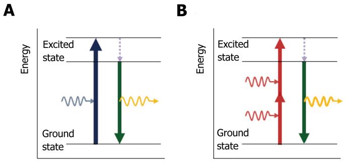
Figure 1 Multiphoton excitation.
A: Single-photon excitation. Individual photons of high-energy blue light (wavelength, λ = 480 nm) excite fluorophores in the sample. After an electron in the fluorophore is transferred to the excited state (blue arrow), it loses energy rapidly owing to non-radiative relaxation (dashed arrow). Subsequently, fluorescence emission (yellow curved arrow) occurs at a longer wavelength than the excitation light as the electron falls back to the ground state (green arrow); B: Two-photon excitation. Two infrared photons (λ = 780 nm) are absorbed simultaneously (red arrows) to excite the fluorophore and light is emitted in the same manner as for single-photon excitation (green arrow) with emission of fluorescein.
- Citation: Cho HJ, Chun HJ, Kim ES, Cho BR. Multiphoton microscopy: An introduction to gastroenterologists. World J Gastroenterol 2011; 17(40): 4456-4460
- URL: https://www.wjgnet.com/1007-9327/full/v17/i40/4456.htm
- DOI: https://dx.doi.org/10.3748/wjg.v17.i40.4456


