Copyright
©2011 Baishideng Publishing Group Co.
World J Gastroenterol. Jan 28, 2011; 17(4): 526-533
Published online Jan 28, 2011. doi: 10.3748/wjg.v17.i4.526
Published online Jan 28, 2011. doi: 10.3748/wjg.v17.i4.526
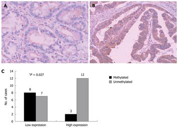
Figure 4 Immunohistochemical analysis of high in normal-1 protein expression in gastric cancer tissue samples.
A: Tumor cells with methylated alleles of high in normal-1 (HIN-1) gene promoter exhibited negative staining; B: Cancer cells without HIN-1 gene promoter methylation exhibited positive staining. HIN-1 expression in gastric cancer (membrane staining, arrow); C: The association of HIN-1 methylation with HIN-1 expression level was analyzed in 29 gastric cancers. High expression: +-+++ staining intensity with 10% or more cancer cells positively stained, otherwise it is considered as low expression. The staining intensity and percentage of staining were compared with a non-cancerous area of the same section. aPearson χ2 test or Pearson χ2 test with continuity correction by SPSS 13.0 software. A, B: IHC, × 200.
- Citation: Gong Y, Guo MZ, Ye ZJ, Zhang XL, Zhao YL, Yang YS. Silence of HIN-1 expression through methylation of its gene promoter in gastric cancer. World J Gastroenterol 2011; 17(4): 526-533
- URL: https://www.wjgnet.com/1007-9327/full/v17/i4/526.htm
- DOI: https://dx.doi.org/10.3748/wjg.v17.i4.526


