Copyright
©2011 Baishideng Publishing Group Co.
World J Gastroenterol. Sep 21, 2011; 17(35): 4048-4051
Published online Sep 21, 2011. doi: 10.3748/wjg.v17.i35.4048
Published online Sep 21, 2011. doi: 10.3748/wjg.v17.i35.4048
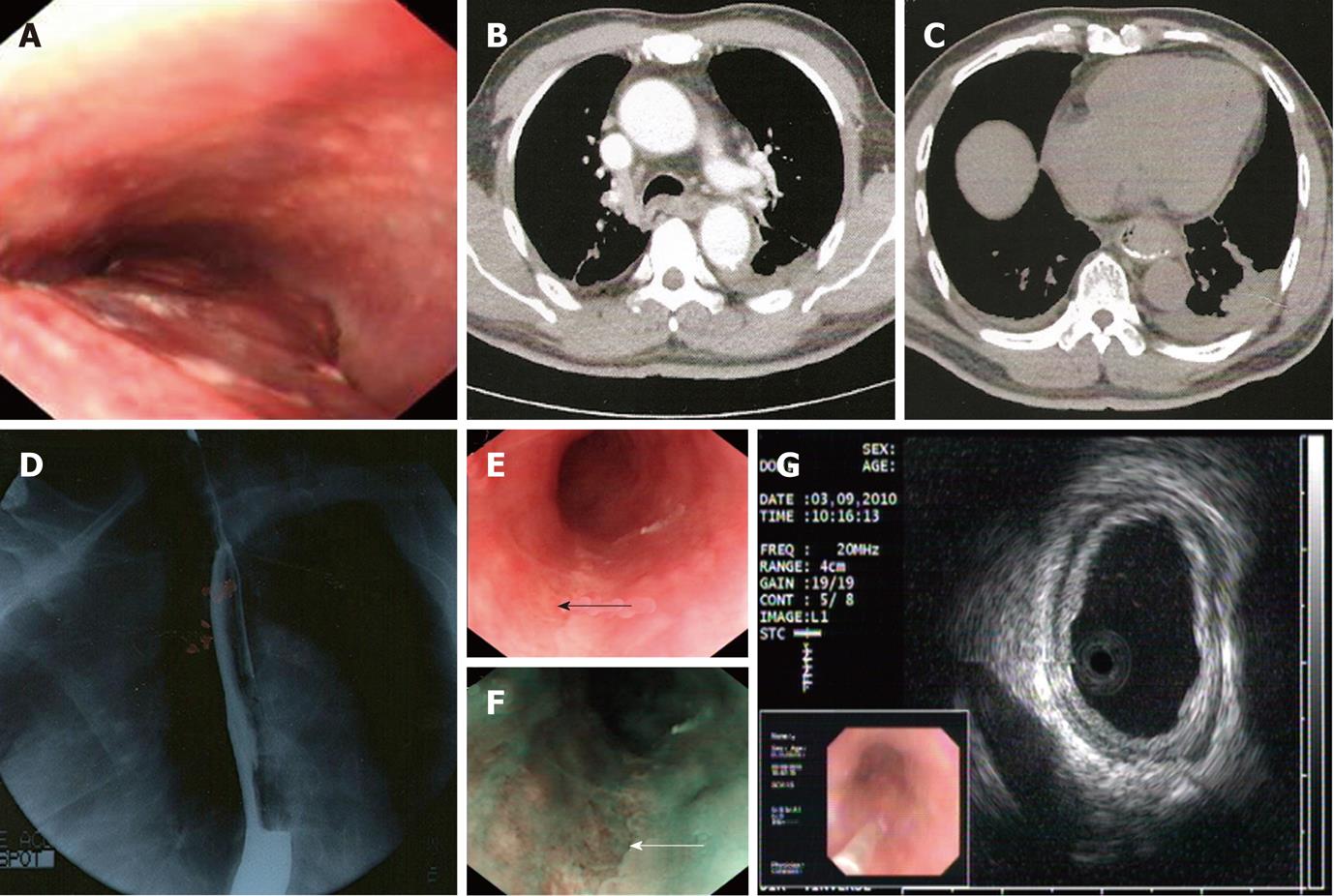
Figure 1 Auxiliary examination.
A: Gastroendoscopy showing edematous mucosa with blood clots; B and C: Obstructive esophageal lumen and lymph nodes with some pleural effusion shown by chest contrast computed tomography; D: Barium X-ray displaying irregular esophageal wall; E and F: The mucosal lesion evaluated by white light imaging (black arrow) or narrow band imaging (white arrow) mode of endoscopy; G: Endoscopic ultrasonography showing focal minimal higher echo.
- Citation: Ma GF, Gao H, Chen SY. Esophageal mucosal lesion with low-dose aspirin and prasugrel mimics malignancy: A case report. World J Gastroenterol 2011; 17(35): 4048-4051
- URL: https://www.wjgnet.com/1007-9327/full/v17/i35/4048.htm
- DOI: https://dx.doi.org/10.3748/wjg.v17.i35.4048


