Copyright
©2011 Baishideng Publishing Group Co.
World J Gastroenterol. Sep 21, 2011; 17(35): 3994-4000
Published online Sep 21, 2011. doi: 10.3748/wjg.v17.i35.3994
Published online Sep 21, 2011. doi: 10.3748/wjg.v17.i35.3994
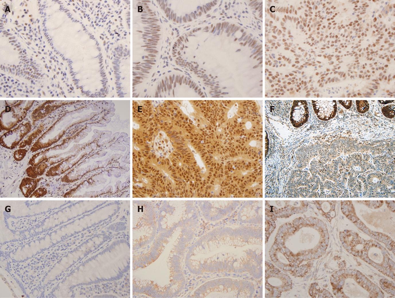
Figure 1 Semi-quantitative immunohistochemistry scoring of transcriptional intermediary factor 1 gamma, Smad4 and transforming growth factor-beta receptor typeII overexpression.
Immunohistochemistry (IHC) staining for transforming growth factor-beta receptor typeII shown in A, B, C: Weak cytoplasmic (0-1+) staining seen in normal colonic mucosa (A). Moderate (2+) cytoplasmic and weak (0-1+) membranous staining in the tubular adenoma (TA) (B). Strong cytoplasmic and focal membranous staining in the cancer cells (C). IHC staining for Smad4 shown in D, E, F: Strong (3+) Smad4 staining seen in normal colonic mucosa (D). Tumour cells showing strong (3+) expression of Smad4 protein in the nucleus and cytoplasm (E). Adenocarcinoma with loss of Smad4 in the nuclei (F). IHC staining for transcriptional intermediary factor 1 gamma (TIF1γ) shown in G, H, I: Weak (1+) nuclear staining for TIF1γ seen in normal colonic mucosa (G), moderate (2+) nuclear staining seen in TA (H), strong nuclear staining (3+) in cancer cells (I) (400 ×).
- Citation: Jain S, Singhal S, Francis F, Hajdu C, Wang JH, Suriawinata A, Wang YQ, Zhang M, Weinshel EH, Francois F, Pei ZH, Lee P, Xu RL. Association of overexpression of TIF1γ with colorectal carcinogenesis and advanced colorectal adenocarcinoma. World J Gastroenterol 2011; 17(35): 3994-4000
- URL: https://www.wjgnet.com/1007-9327/full/v17/i35/3994.htm
- DOI: https://dx.doi.org/10.3748/wjg.v17.i35.3994


