Copyright
©2011 Baishideng Publishing Group Co.
World J Gastroenterol. Sep 7, 2011; 17(33): 3850-3855
Published online Sep 7, 2011. doi: 10.3748/wjg.v17.i33.3850
Published online Sep 7, 2011. doi: 10.3748/wjg.v17.i33.3850
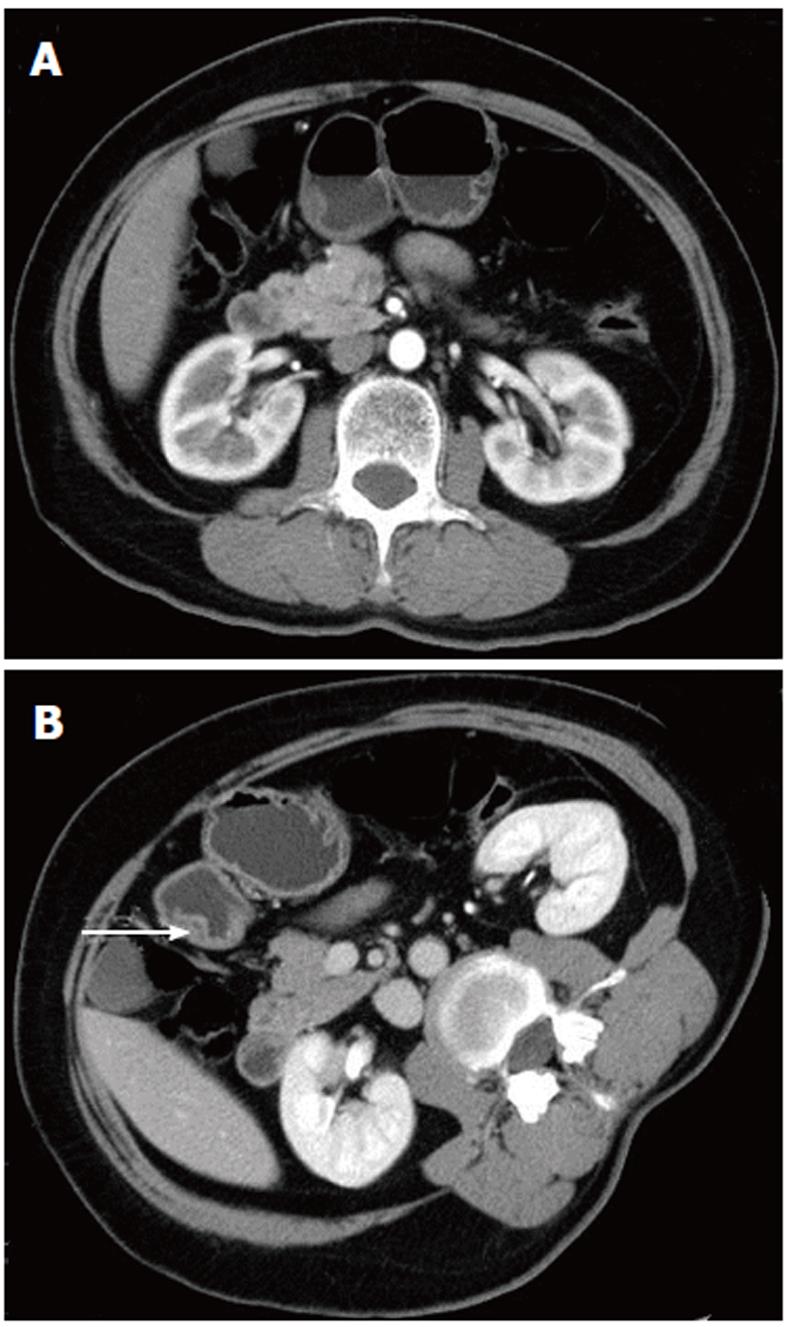
Figure 1 Heterotopic pancreas in the gastric antrum of a 59-year-old woman.
A: Arterial phase image showing gastric antrum wall thickening. There is a circumscribed border of the lesion below the mucosal layer. The density at the center of the lesion is slightly lower than at the periphery. B: Each patient was asked to lie on their right side during venous phase scanning. The central area of the low-density region (white arrow) is obvious.
- Citation: Wang D, Wei XE, Yan L, Zhang YZ, Li WB. Enhanced CT and CT virtual endoscopy in diagnosis of heterotopic pancreas. World J Gastroenterol 2011; 17(33): 3850-3855
- URL: https://www.wjgnet.com/1007-9327/full/v17/i33/3850.htm
- DOI: https://dx.doi.org/10.3748/wjg.v17.i33.3850


