Copyright
©2011 Baishideng Publishing Group Co.
World J Gastroenterol. Aug 28, 2011; 17(32): 3733-3738
Published online Aug 28, 2011. doi: 10.3748/wjg.v17.i32.3733
Published online Aug 28, 2011. doi: 10.3748/wjg.v17.i32.3733
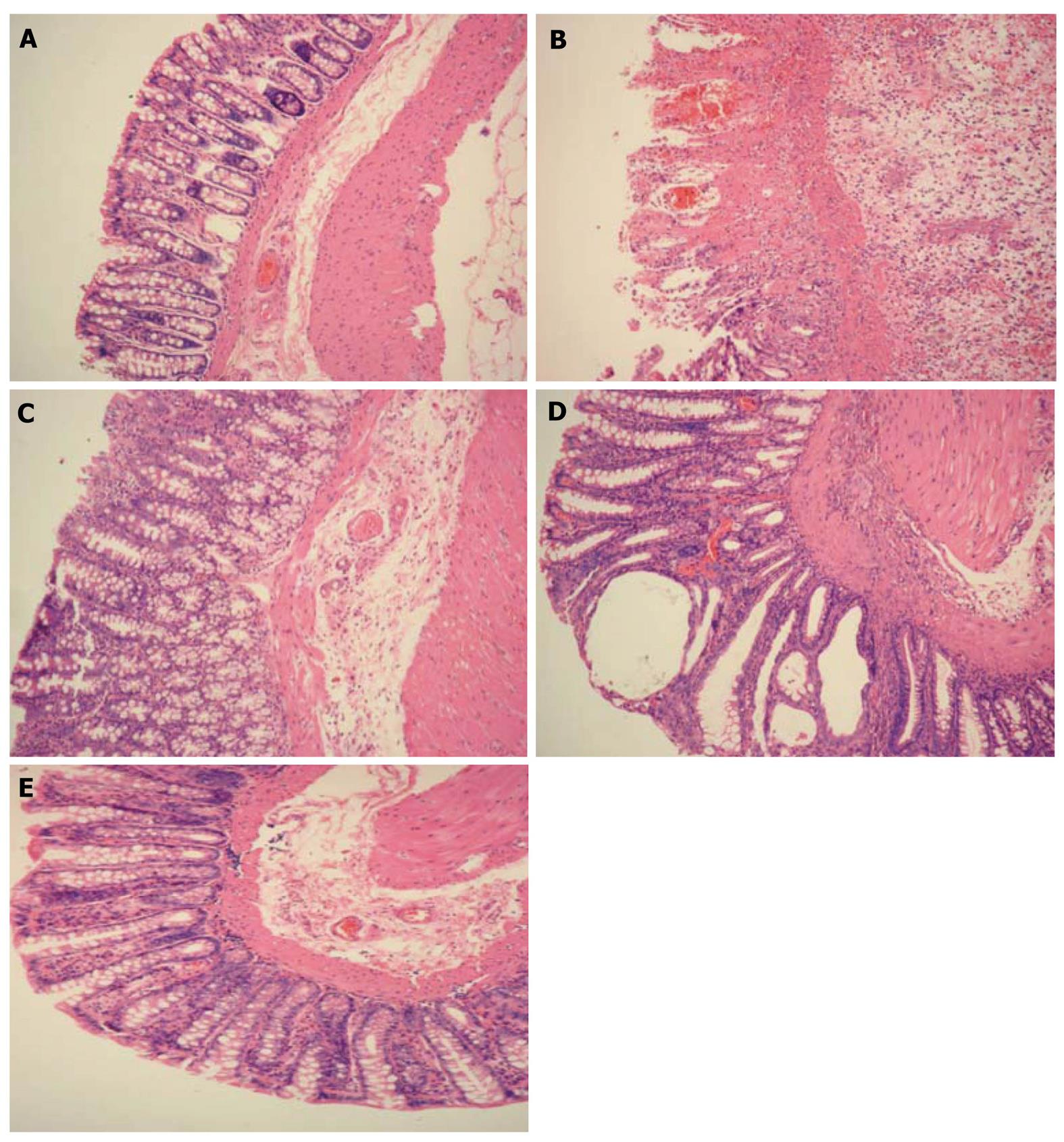
Figure 1 Results of hematoxylin eosine staining of rat colonic tissue (× 100).
A: Normal group: the colonic mucosa was complete and the colonic gland was regularly arranged, with no apparent inflammatory cell infiltration; B: Model group: damage to colonic mucosa was found and there were monocytes and a large number of inflammatory cells infiltrating the mucosa or submucosa; C: Moxibustion group: the colonic gland was regularly arranged compared with the model group and ulceration was covered by regenerated epithelium; D: Moxibustion plus disodium cromoglycate group: slight congestion of colonic mucosa and fibroplasia of submucosa were found and a large number of infiltrating inflammatory cells, however, this was not as serious as in the model group; E: Moxibustion plus normal saline group: the colonic gland was regularly arranged and inflammatory cell infiltration of the submucosa was noted.
- Citation: Shi Y, Qi L, Wang J, Xu MS, Zhang D, Wu LY, Wu HG. Moxibustion activates mast cell degranulation at the ST25 in rats with colitis. World J Gastroenterol 2011; 17(32): 3733-3738
- URL: https://www.wjgnet.com/1007-9327/full/v17/i32/3733.htm
- DOI: https://dx.doi.org/10.3748/wjg.v17.i32.3733


