Copyright
©2011 Baishideng Publishing Group Co.
World J Gastroenterol. Aug 21, 2011; 17(31): 3614-3622
Published online Aug 21, 2011. doi: 10.3748/wjg.v17.i31.3614
Published online Aug 21, 2011. doi: 10.3748/wjg.v17.i31.3614
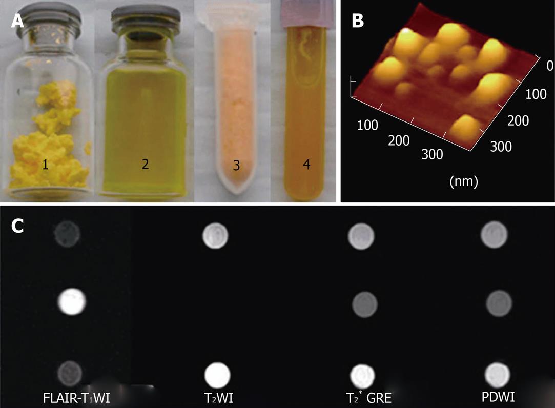
Figure 1 Characterization of solid lipid nanoparticles.
A: Gadopentetate dimeglumine and fluorescein isothiocyanate-loaded solid lipid nanoparticles (Gd-FITC-SLNs) freeze-dried powder (1) and dispersion (2); Fluorescein isothiocyanate solid lipid nanoparticles (FITC-SLNs) freeze-dried powder (3) and dispersion (4); B: Atomic force microscopy images of blank solid lipid nanoparticles; C: Magnetic resonance (MR) images of FITC-SLNs dispersions (top). Gd-FITC-SLN suspension (middle). Water (bottom) obtained with fluid attenuated inversion recovery (FLAIR) (left ), T2WI (middle left ), T2* GRE (middle right) and PDWI (right). FLAIR was obtained with the following parameters: 2000/11.1/750/2, TR/TE/TI/NEX; T2WI: 3860/106/2 (TR/TE/NEX); T2* GRE: 550/14/2/200 (TR/TE/NEX/Flip); PDWI: 3220/12/1 (TR/TE/NEX). Both sequences used a 256 × 160 matrix, a 140 mm FOV, and 4-mm-thick sections.
- Citation: Wu T, Zheng WL, Zhang SZ, Sun JH, Yuan H. Bimodal visualization of colorectal uptake of nanoparticles in dimethylhydrazine-treated mice. World J Gastroenterol 2011; 17(31): 3614-3622
- URL: https://www.wjgnet.com/1007-9327/full/v17/i31/3614.htm
- DOI: https://dx.doi.org/10.3748/wjg.v17.i31.3614


