Copyright
©2011 Baishideng Publishing Group Co.
World J Gastroenterol. Aug 21, 2011; 17(31): 3596-3604
Published online Aug 21, 2011. doi: 10.3748/wjg.v17.i31.3596
Published online Aug 21, 2011. doi: 10.3748/wjg.v17.i31.3596
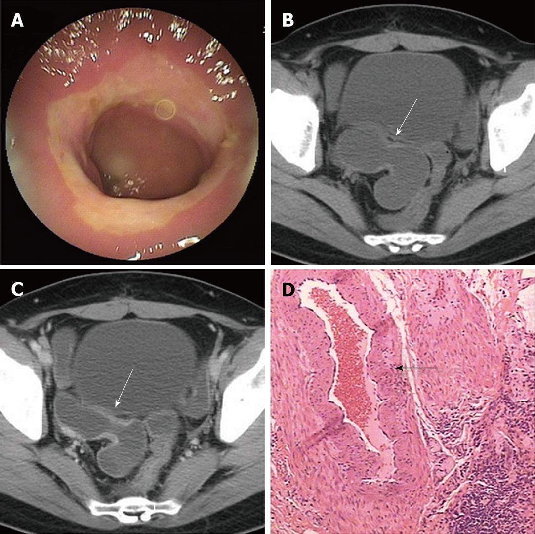
Figure 3 Forty-four-year-old woman presenting with a 4-year history of recurrent episodes of hematochezia and abdominal pain.
A: Double-balloon enteroscopy image showing circumferential stricture in the ileum; B: Computed tomography (CT) plain scan image showing mild bowel wall thickening and circumferential stricture of the lumen in the ileum (arrow); C: Contrast-enhanced CT scan image showing that contrast enhancement of thickened bowel wall is homogenous and moderate (arrow); D: Blood vessel with thickened wall and expanded lumen shown in the submucosa (hematoxylin and eosin, orig. mag × 40).
- Citation: Wang ML, Miao F, Tang YH, Zhao XS, Zhong J, Yuan F. Special diaphragm-like strictures of small bowel unrelated to non-steroidal anti-inflammatory drugs. World J Gastroenterol 2011; 17(31): 3596-3604
- URL: https://www.wjgnet.com/1007-9327/full/v17/i31/3596.htm
- DOI: https://dx.doi.org/10.3748/wjg.v17.i31.3596


