Copyright
©2011 Baishideng Publishing Group Co.
World J Gastroenterol. Aug 14, 2011; 17(30): 3544-3553
Published online Aug 14, 2011. doi: 10.3748/wjg.v17.i30.3544
Published online Aug 14, 2011. doi: 10.3748/wjg.v17.i30.3544
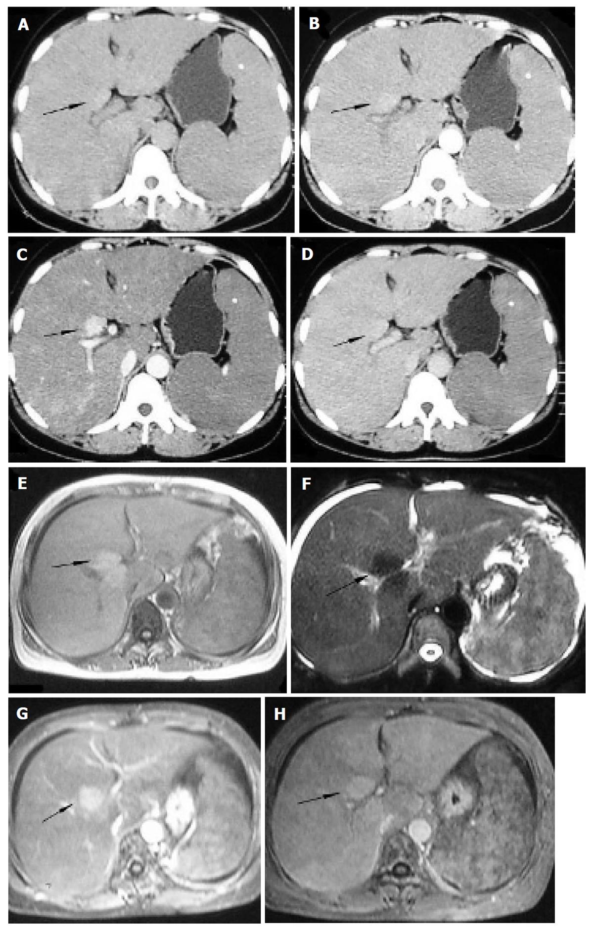
Figure 4 Diffuse hepatic epithelioid hemangioendothelioma in a 48-year-old woman.
Plain computed tomography and magnetic resonance imaging manifests an obviously enlarged liver with a large nodule (black arrow) appearing slightly hyperdense relative to normal liver parenchyma (A), isointense on T1WI (E) and hypointense on T2WI (F), with slight enhancement in the arterial phase (B), evident enhancement in the portal venous phase (C and G) and iso-density/intensity in the equilibrium phase (D and H), associated with splenomegaly and ascites (F).
- Citation: Chen Y, Yu RS, Qiu LL, Jiang DY, Tan YB, Fu YB. Contrast-enhanced multiple-phase imaging features in hepatic epithelioid hemangioendothelioma. World J Gastroenterol 2011; 17(30): 3544-3553
- URL: https://www.wjgnet.com/1007-9327/full/v17/i30/3544.htm
- DOI: https://dx.doi.org/10.3748/wjg.v17.i30.3544


