Copyright
©2011 Baishideng Publishing Group Co.
World J Gastroenterol. Aug 14, 2011; 17(30): 3503-3509
Published online Aug 14, 2011. doi: 10.3748/wjg.v17.i30.3503
Published online Aug 14, 2011. doi: 10.3748/wjg.v17.i30.3503
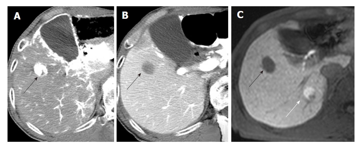
Figure 3 An 80-year-old man with poorly differentiated (black arrow) and well differentiated hepatocellular carcinoma (white arrow).
A: Poorly differentiated hepatocellular carcinoma shows hypervascularity on computed tomography (CT) hepatic arteriography; B: Hypodensity on CT during arterial portography; C: Hypointensity in hepatobiliary phase on gadoxetic acid-enhanced magnetic resonance imaging (MRI). The lesion is not visible on CT hepatic arteriography (A) or on CT during arterial portography (B) and shows hyperintensity in the hepatobiliary phase on gadoxetic acid-enhanced MRI.
- Citation: Saito K, Moriyasu F, Sugimoto K, Nishio R, Saguchi T, Nagao T, Taira J, Akata S, Tokuuye K. Diagnostic efficacy of gadoxetic acid-enhanced MRI for hepatocellular carcinoma and dysplastic nodule. World J Gastroenterol 2011; 17(30): 3503-3509
- URL: https://www.wjgnet.com/1007-9327/full/v17/i30/3503.htm
- DOI: https://dx.doi.org/10.3748/wjg.v17.i30.3503


