Copyright
©2011 Baishideng Publishing Group Co.
World J Gastroenterol. Jan 21, 2011; 17(3): 300-312
Published online Jan 21, 2011. doi: 10.3748/wjg.v17.i3.300
Published online Jan 21, 2011. doi: 10.3748/wjg.v17.i3.300
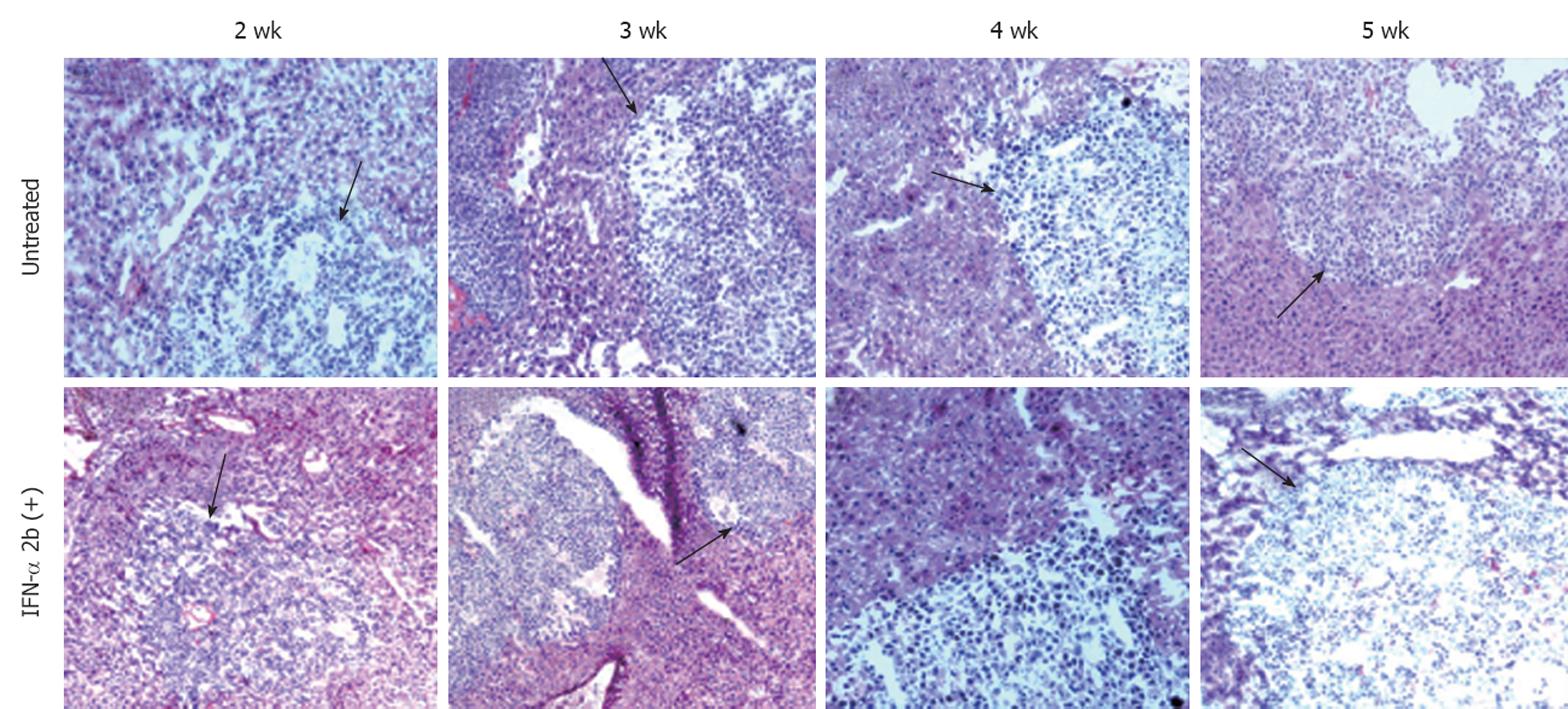
Figure 9 Histological evaluation of liver sections after hematoxylin and eosin staining that indicates that interferon-α treatment did not cause any tumor necrosis or reduce the size of tumor nodules in the SCID mice liver.
The upper panel shows the untreated mouse liver sections after 2-5 wk of tumor development. The black arrows indicate the hepatocellular carcinoma (HCC) tumor in the mouse liver. Similarly, the lower panel shows the interferon-α-treated mouse liver sections with HCC tumor at different time points (10 × magnification).
- Citation: Hazari S, Hefler HJ, Chandra PK, Poat B, Gunduz F, Ooms T, Wu T, Balart LA, Dash S. Hepatocellular carcinoma xenograft supports HCV replication: A mouse model for evaluating antivirals. World J Gastroenterol 2011; 17(3): 300-312
- URL: https://www.wjgnet.com/1007-9327/full/v17/i3/300.htm
- DOI: https://dx.doi.org/10.3748/wjg.v17.i3.300


