Copyright
©2011 Baishideng Publishing Group Co.
World J Gastroenterol. Jan 21, 2011; 17(3): 300-312
Published online Jan 21, 2011. doi: 10.3748/wjg.v17.i3.300
Published online Jan 21, 2011. doi: 10.3748/wjg.v17.i3.300
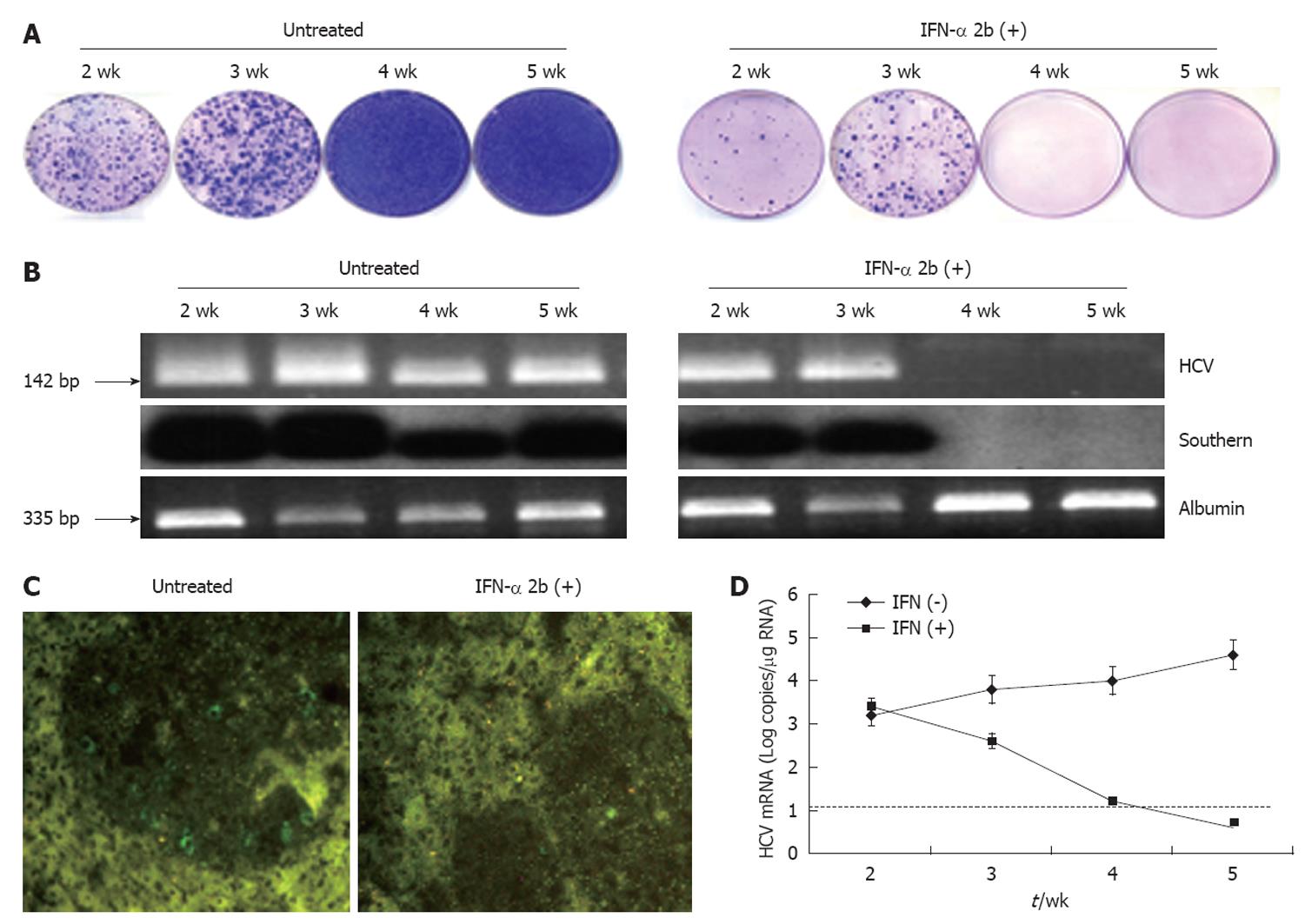
Figure 8 Antiviral effect of interferon-α in the liver tumor model.
S3-green fluorescence protein (GFP) cells were implanted in the liver of NOD/SCID mice by intrasplenic injection. After 3 wk, interferon-α (IFN-α) treatment was started. The time shown represents the weeks after IFN treatment. A: Colony assay of viable tumor cells isolated from the liver tumor model at different time points with or without IFN-α treatment. Tumor cells isolated from the entire mouse liver were cultured in the medium that contained 1 mg/mL G-418. The replicon cells with hepatitis C virus (HCV) survived the treatment and formed cell colonies. Left panel: there was an increase in the number of cell colonies between 4 and 5 wk. Right panel: IFN-α treatment completely inhibited HCV replication and cell colony formation at 4 wk; B: RT-nested polymerase chain reaction (PCR) and Southern blot analysis for HCV and albumin in the RNA extracts of the liver tumor. Left panel shows the results without treatment. Right panel shows IFN-α treatment; C: Expression of HCV-GFP in the liver tumors before and after IFN treatment. IFN treatment after 4 wk completely inhibited HCV-GFP expression in the S3-GFP tumors in the SCID mouse liver; D: Real-time reverse transcription PCR (RT-PCR) showed that the levels of HCV in the liver tumor remained undetected after 4 wk of IFN-α treatment. The dotted line indicates the limit of detection for real time RT-PCR assay.
- Citation: Hazari S, Hefler HJ, Chandra PK, Poat B, Gunduz F, Ooms T, Wu T, Balart LA, Dash S. Hepatocellular carcinoma xenograft supports HCV replication: A mouse model for evaluating antivirals. World J Gastroenterol 2011; 17(3): 300-312
- URL: https://www.wjgnet.com/1007-9327/full/v17/i3/300.htm
- DOI: https://dx.doi.org/10.3748/wjg.v17.i3.300


