Copyright
©2011 Baishideng Publishing Group Co.
World J Gastroenterol. Jan 21, 2011; 17(3): 300-312
Published online Jan 21, 2011. doi: 10.3748/wjg.v17.i3.300
Published online Jan 21, 2011. doi: 10.3748/wjg.v17.i3.300
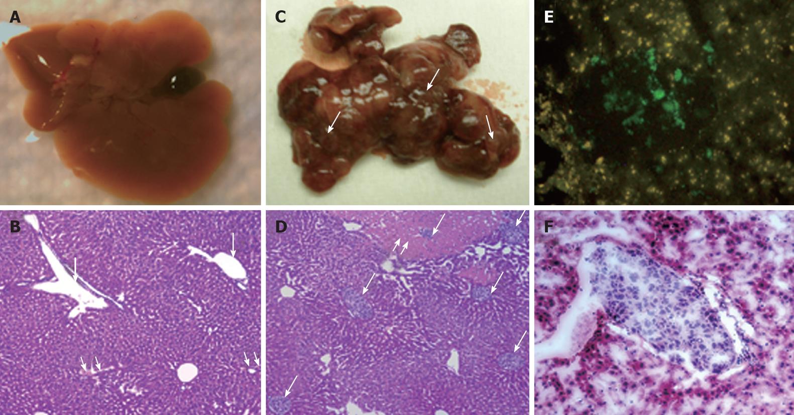
Figure 3 Human hepatocellular carcinoma xenograft in SCID/NOD mouse liver.
A: Gross appearance of normal mouse liver with distended gall bladder; B: Light microscopic appearance of normal liver stained with hematoxylin and eosin (4 × magnification). The portal tracts are shown by single arrows and the central veins are marked with double arrows; C: Gross appearance of mouse liver with metastatic nodules of hepatocellular carcinoma (HCC). Note the distended white-tan areas of metastasis and infarction in the mouse liver (single arrows); D: Microscopic metastasis of HCC diffusely infiltrating the liver through the portal venous system (single arrows). Note the areas of infarction secondary to the tumor emboli in the portal vein (double arrow). Human hepatocytes can be easily discriminated from the mouse hepatocytes by their size and pale color; E: Expression of hepatitis C virus-green fluorescence protein (GFP) fusion protein in the S3-GFP liver tumors in the mouse; F: Hematoxylin and eosin staining (frozen section) of HCC tumor in the mouse liver at 4 wk after intrasplenic infusion of S3-GFP replicon cells.
- Citation: Hazari S, Hefler HJ, Chandra PK, Poat B, Gunduz F, Ooms T, Wu T, Balart LA, Dash S. Hepatocellular carcinoma xenograft supports HCV replication: A mouse model for evaluating antivirals. World J Gastroenterol 2011; 17(3): 300-312
- URL: https://www.wjgnet.com/1007-9327/full/v17/i3/300.htm
- DOI: https://dx.doi.org/10.3748/wjg.v17.i3.300


