Copyright
©2011 Baishideng Publishing Group Co.
World J Gastroenterol. Aug 7, 2011; 17(29): 3459-3464
Published online Aug 7, 2011. doi: 10.3748/wjg.v17.i29.3459
Published online Aug 7, 2011. doi: 10.3748/wjg.v17.i29.3459
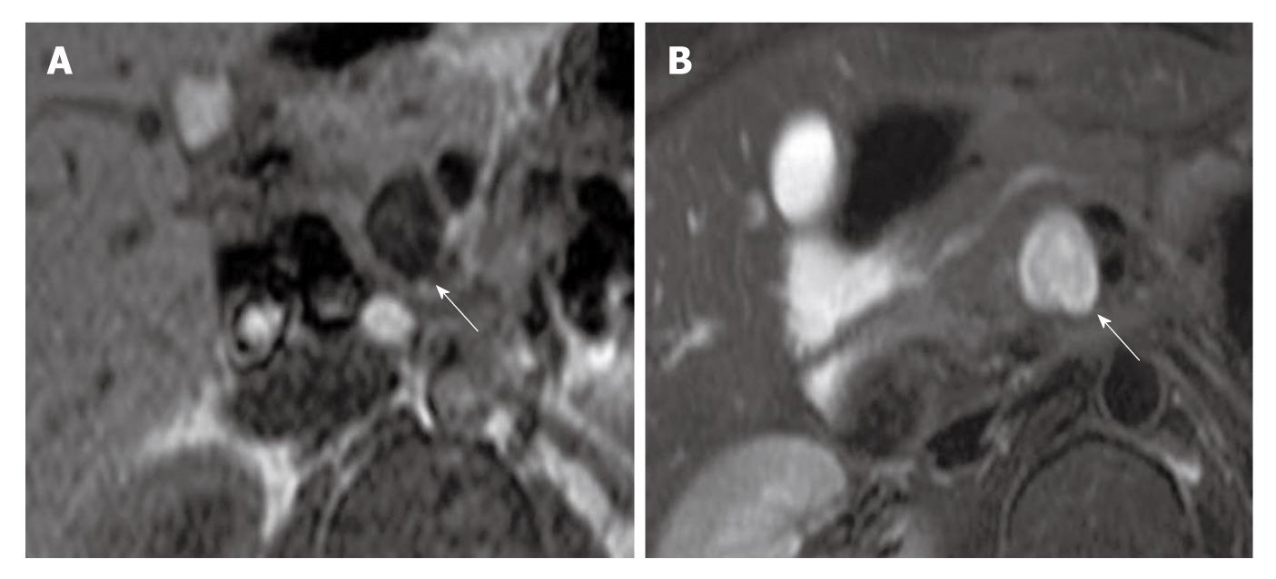
Figure 4 Magnetic resonance imaging of Case 1 showed a round mass (arrows) with low signal intensity in T1-weighted images and high signal intensity in T2-weighted images.
The tumor signal was uniform. A: T1-weighted image; B: T2-weighted image.
- Citation: Kudo T, Kawakami H, Kuwatani M, Ehira N, Yamato H, Eto K, Kubota K, Asaka M. Three cases of retroperitoneal schwannoma diagnosed by EUS-FNA. World J Gastroenterol 2011; 17(29): 3459-3464
- URL: https://www.wjgnet.com/1007-9327/full/v17/i29/3459.htm
- DOI: https://dx.doi.org/10.3748/wjg.v17.i29.3459


