Copyright
©2011 Baishideng Publishing Group Co.
World J Gastroenterol. Jul 28, 2011; 17(28): 3335-3341
Published online Jul 28, 2011. doi: 10.3748/wjg.v17.i28.3335
Published online Jul 28, 2011. doi: 10.3748/wjg.v17.i28.3335
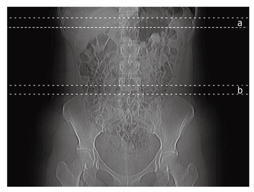
Figure 1 Scanning range of the liver and abdominal fat.
Seven serial axial slices with a thickness of 3 mm were scanned for the liver and the spleen. Another seven slices from the iliac crest and above were scanned to assess abdominal fat. a: The range for hepatic fat examination; b: The range for abdominal fat examination.
- Citation: Jang S, Lee CH, Choi KM, Lee J, Choi JW, Kim KA, Park CM. Correlation of fatty liver and abdominal fat distribution using a simple fat computed tomography protocol. World J Gastroenterol 2011; 17(28): 3335-3341
- URL: https://www.wjgnet.com/1007-9327/full/v17/i28/3335.htm
- DOI: https://dx.doi.org/10.3748/wjg.v17.i28.3335


