Copyright
©2011 Baishideng Publishing Group Co.
World J Gastroenterol. Jul 28, 2011; 17(28): 3300-3309
Published online Jul 28, 2011. doi: 10.3748/wjg.v17.i28.3300
Published online Jul 28, 2011. doi: 10.3748/wjg.v17.i28.3300
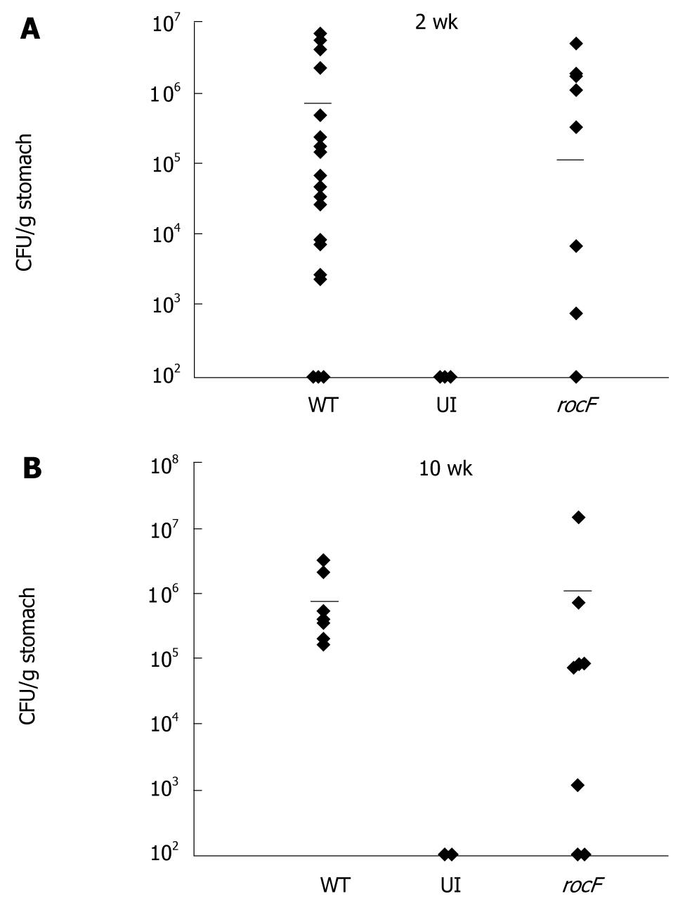
Figure 4 The rocF mutant of Helicobacter pylori colonizes the stomach of arginase II knockout mice.
Arginase II knockout mice were inoculated with either the wild-type SS1 strain (WT) of Helicobacter pylori (H. pylori) or the isogenic rocF mutant (rocF). Number of animals used per group were as follows: 2 wk experiment: 13 for WT, nine for rocF mutant; 10 wk experiment: eight for WT, eight for rocF mutant. At 2 (A) or 10 (B) wk post-infection, stomachs were removed, completely homogenized, and plated for H. pylori. UI, uninfected controls (n = 2 at 2 and 10 wk). Limit of detection: ~100 CFU/g stomach; all animals that lacked H. pylori were set to this detection limit. Bar, mean CFU/g stomach. At 2 wk post-infection, P = 0.65 by unpaired two-tailed t test between the mean CFU/g tissue for the wild-type versus the rocF mutant. Each symbol represents one mouse. In some cases there are two mice represented by a symbol if the data overlapped.
-
Citation: Kim SH, Langford ML, Boucher JL, Testerman TL, McGee DJ.
Helicobacter pylori arginase mutant colonizes arginase II knockout mice. World J Gastroenterol 2011; 17(28): 3300-3309 - URL: https://www.wjgnet.com/1007-9327/full/v17/i28/3300.htm
- DOI: https://dx.doi.org/10.3748/wjg.v17.i28.3300


