Copyright
©2011 Baishideng Publishing Group Co.
World J Gastroenterol. Jun 28, 2011; 17(24): 2972-2975
Published online Jun 28, 2011. doi: 10.3748/wjg.v17.i24.2972
Published online Jun 28, 2011. doi: 10.3748/wjg.v17.i24.2972
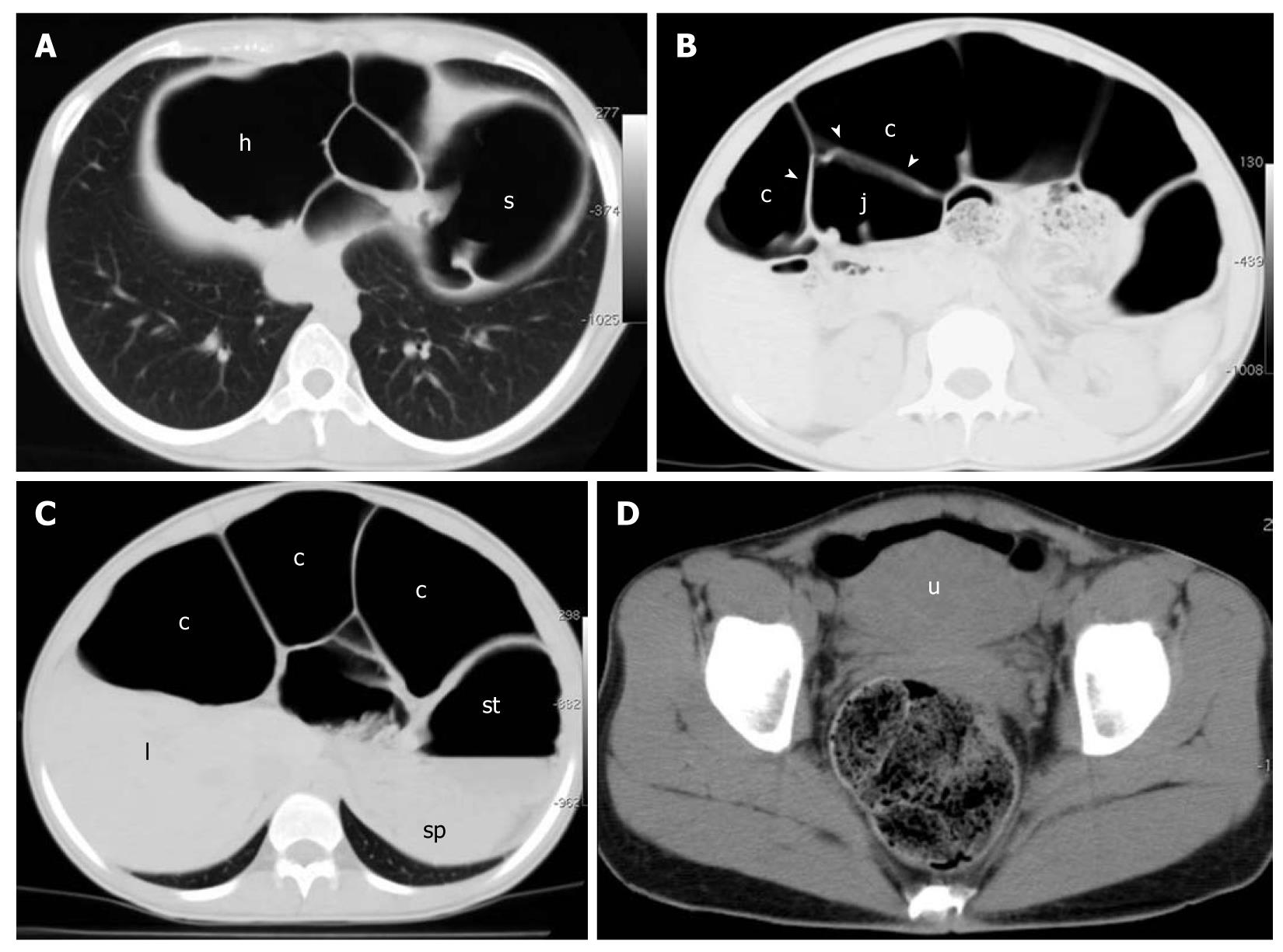
Figure 2 Unenhanced four-row MDCT.
Images are displayed with lung (A-C) and soft tissue (D) windows. A: Hepatic (h) and splenic (s) flexures, air-filled and abnormally dilated, are displaced under the diaphragm, accounting for the subphrenic radiolucency depicted in the upright film; B: Intraluminal air in the large bowel (c) appears to outline the intestinal wall (arrowheads) of a jejunal loop (j) that is also mildly dilated and air-filled, accounting for the Rigler’s sign depicted on the upright and supine films; C: Abnormally dilated colonic segment situated between the liver (l) and anterior abdominal wall, simulating the hyperlucent (bright) liver sign depicted in the supine film; st: Stomach; sp: Spleen; D: Rectum is normally filled with feces. u: Uterus.
- Citation: Camera L, Calabrese M, Sarnelli G, Longobardi M, Rocco A, Cuomo R, Salvatore M. Pseudopneumoperitoneum in chronic intestinal pseudo-obstruction: A case report. World J Gastroenterol 2011; 17(24): 2972-2975
- URL: https://www.wjgnet.com/1007-9327/full/v17/i24/2972.htm
- DOI: https://dx.doi.org/10.3748/wjg.v17.i24.2972


